Cardiovascular system · Head and neck
Circle of Willis !
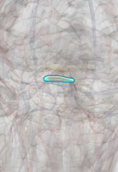
The circle of Willis is a ring of arteries under the brain that links its main blood supplies and can reroute blood; aneurysms form at its junctions.
Official name: Cerebral arterial circle
Opens in the Durgical app, no sign-up needed.
What it is
The ring of arteries under the brain linking the carotid and vertebral systems.
What it does
Can reroute blood if one artery narrows; aneurysms form at its junctions.
Key facts
- Origin
- Both internal carotid arteries and the basilar artery
- Branches
- Anterior cerebral arteries + anterior communicating, internal carotids, posterior communicating arteries, posterior cerebral arteries
- Supplies
- Whole brain via the cerebral arteries; collateral route between sides; often incomplete; aneurysms form at its junctions
Relations
- Contents
- Encircles the optic chiasm, the pituitary stalk (infundibulum), the tuber cinereum, the mammillary bodies and the posterior perforated substance
- Above
- Base of the brain: the hypothalamus and the inferior surfaces of the frontal and temporal lobes
- Below
- Sella turcica with the pituitary gland under its diaphragm; the circle lies in the subarachnoid cisterns at the base of the skull
- Behind
- Interpeduncular fossa of the midbrain, where the oculomotor nerves emerge between the posterior cerebral and superior cerebellar arteries
How to find it
It lies at the base of the brain in the cisterns around the optic chiasm, the pituitary stalk and the mammillary bodies, under the hypothalamus. On axial CT or MR angiography it is seen as a ring in the suprasellar region; surgically, its front half is reached through a pterional craniotomy.
On imaging
Time-of-flight MR angiography shows it without contrast; CT angiography is the fastest test in an emergency; catheter angiography gives the finest detail when planning aneurysm treatment.
Clinical notes
- When one internal carotid narrows slowly, a complete circle can keep the brain supplied, so an occlusion may cause no symptoms; an incomplete circle protects less
- A posterior communicating aneurysm can press on the oculomotor nerve: drooping lid, eye turned down and out, and a dilated pupil
- Pituitary tumours growing up out of the sella lie within the circle and can reach its vessels and the optic chiasm
- Days after a subarachnoid haemorrhage, spasm of the circle's arteries (vasospasm) can cause delayed ischaemia
- Two separate systems leave the circle: short central (perforating) end arteries to the basal ganglia, thalamus and internal capsule, and cortical branches that join over the surface through pial anastomoses; these collaterals can rescue the cortex after a branch occlusion but not the deep nuclei
Anatomical variants
- Fetal-type posterior cerebral artery, filled from the internal carotid through a large posterior communicating artery, with a small P1 segment
- Small or absent A1 segment, so both anterior cerebral arteries fill from one side
- Duplicated or fenestrated anterior communicating artery
Development
Each internal carotid first splits into a front branch (ophthalmic, anterior and middle cerebral) and a back branch that later joins the vertebral system; how much of the posterior circulation the carotid keeps explains the fetal-type posterior cerebral artery.
Related conditions
- Subarachnoid haemorrhage
- Brain arteriovenous malformation
- Primary angiitis of the central nervous system
Test yourself
Think of the answer, then tap the question to see it.
Which arteries form the circle of Willis?
Both internal carotids, the anterior cerebral and anterior communicating arteries, the posterior communicating and posterior cerebral arteries (from the basilar).
Where do berry aneurysms of the circle of Willis form, in order of frequency?
At branch points: anterior communicating artery, internal carotid–posterior communicating junction, middle cerebral bifurcation, basilar tip.
How does a ruptured aneurysm of the circle of Willis present?
As subarachnoid haemorrhage: sudden thunderclap headache, the worst ever, often with neck stiffness, vomiting or collapse.
What is the work-up for suspected subarachnoid haemorrhage?
Non-contrast CT first, then CT angiography; if CT is normal but suspicion is high, lumbar puncture for xanthochromia.
How common is a complete, balanced circle of Willis?
Found in a minority (under half in many series); small posterior communicating arteries and A1 segments are the usual gaps.
Educational information, not medical advice. For any symptom, see a health professional.
Its names
- Official Latin (TA2)
- circulus arteriosus cerebri TA2 4516
- Official Spanish (DeCS)
- Círculo Arterial Cerebral DeCS D002941
- English (TA2)
- Cerebral arterial circle
- Also called
- circulus Willisi
Sources: Terminologia Anatomica (TA2); DeCS; Neuroanatomy, Circle of Willis; Anatomy of the Human Body (20th ed.): Development of the Vascular System (Lea & Febiger (Gray's Anatomy, 1918), 1918) · adapted, Public domain; Tratado de anatomía humana (9.ª ed.), tomo II, Sistema nervioso central: cerebro, núcleos grises centrales, vías de conducción y circulación cerebral.
Spotted a mistake?We are always improving for the community. Tell us what is wrong and our team will review it.Report a mistake
Representative model, for educational purposes only.