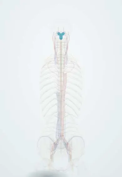
Anterior spinal artery !
The anterior spinal artery is a single small artery down the front of the spinal cord; it feeds the front two-thirds, including movement pathways.
Opens in the Durgical app, no sign-up needed.
What it is
A single small artery running down the front of the spinal cord.
What it does
Supplies the front two-thirds of the cord, including the movement pathways.
Key facts
- Origin
- Union of a branch from each vertebral artery near the foramen magnum
- Branches
- Sulcal (central) arteries into the cord; reinforced along its length by segmental medullary arteries from the vertebral, ascending and deep cervical, posterior intercostal, lumbar and lateral sacral arteries (e.g. Adamkiewicz)
- Supplies
- Anterior two-thirds of the cord: anterior horns, corticospinal and spinothalamic tracts; occlusion spares the dorsal columns
Relations
- Course
- Formed near the foramen magnum by a branch from each vertebral artery, which alone feeds only the upper cervical cord; below, it runs down the anterior median fissure as a chain kept up by a few anterior segmental medullary arteries, and joins the posterior spinal arteries in a loop around the conus medullaris
- Deep
- Sulcal (central) arteries enter the fissure to reach the anterior horns and the anterior and lateral columns
- Lateral
- Anterior nerve rootlets on each side, along which the segmental medullary arteries arrive to join it
- Superficial
- Pia mater, which carries it, then the subarachnoid space, the arachnoid and the dura
Examination
- Comparison of pinprick and temperature with vibration (tuning fork) and joint position: loss of the first two with the last two kept points to the anterior cord
On imaging
MRI with diffusion-weighted sequences shows the infarct; on axial T2 images the bright, paired anterior horns give the "owl's eyes" (or "snake eyes") sign.
Clinical notes
- Classic causes: surgery or stent-grafting of the thoracoabdominal aorta, aortic dissection, severe hypotension and, in the neck, compression by a large disc or osteophyte
- Particulate injections around a nerve root (transforaminal epidural injection) can, rarely, enter a radicular feeder and infarct the cord
- Weakness is often flaccid at first, with spasticity and brisk reflexes developing below the level over the following weeks
Development
It forms when paired longitudinal channels on the front of the cord fuse in the midline; short doubled segments can remain.
Related conditions
Test yourself
Think of the answer, then tap the question to see it.
How is the anterior spinal artery formed?
By the union of a branch from each vertebral artery near the foramen magnum; it then runs down the anterior median fissure.
What deficits define anterior spinal artery syndrome?
Sudden paraplegia or quadriplegia, loss of pain and temperature below the level and bladder dysfunction, with vibration and joint position sense preserved.
Why are vibration and position sense spared when the anterior spinal artery is occluded?
The anterior spinal artery supplies only the anterior two-thirds of the cord; the dorsal columns belong to the posterior spinal arteries.
Which segment of the cord supplied by the anterior spinal artery is most vulnerable to low blood pressure?
The mid-thoracic cord, about T4–T9: feeders are sparse there, making it a watershed zone.
What MRI sign suggests an anterior spinal artery infarct?
The «owl's eyes» (or «snake eyes») sign: bright paired anterior horns on axial T2, with restricted diffusion.
Educational information, not medical advice. For any symptom, see a health professional.
Its names
- Official Latin (TA2)
- arteria spinalis anterior TA2 4525
- Official Spanish (DeCS)
- Arteria espinal anterior
- English (TA2)
- Anterior spinal artery
- Also called
- arteria spinalis ventralis
Related
Sources: Terminologia Anatomica (TA2); Neuroanatomy, Spinal Cord; Tratado de anatomía humana (9.ª ed.), tomo II, Libro V Sistema nervioso central, sección 1: Médula espinal.
Spotted a mistake?We are always improving for the community. Tell us what is wrong and our team will review it.Report a mistake
Representative model, for educational purposes only.