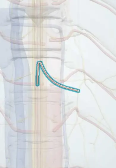
Artery of Adamkiewicz !
The artery of Adamkiewicz is the largest artery feeding the lower spinal cord, usually entering on the left between T8 and L2.
Official name: Great radicular artery
Opens in the Durgical app, no sign-up needed.
What it is
The largest artery feeding the front of the lower spinal cord. It usually enters on the left between T8 and L2, but the level varies.
What it does
Supplies much of the lower two-thirds of the cord through the anterior spinal artery; injury can cause paralysis, so surgeons and radiologists take it into account.
Key facts
- Origin
- Usually a left posterior intercostal or lumbar artery at T9–T12 (about 75%; varies T5–L2)
- Branches
- Great anterior segmental medullary artery; joins the anterior spinal artery with a hairpin turn
- Supplies
- Lower two-thirds of the cord via the anterior spinal artery (lumbosacral enlargement and conus)
Relations
- Course
- From the dorsal (spinal) branch of a lower posterior intercostal or upper lumbar artery, usually on the left, it enters an intervertebral foramen with a spinal nerve, climbs along the ventral root and then turns sharply down to join the anterior spinal artery
How to find it
It cannot be felt. It usually enters the canal on the left through a foramen between T9 and T12, climbs along the root and joins the anterior spinal artery with a sharp downward hairpin turn, the sign used to recognize it on spinal angiography or CT angiography.
On imaging
Spinal angiography with selective injection of the intercostal and lumbar arteries is the reference; CT or MR angiography can often show it without a catheter as a thin vessel on the front of the cord with a hairpin turn.
Clinical notes
- Paraplegia after thoracoabdominal aneurysm repair or stent-grafting reflects lost flow through this artery and its collaterals; reimplanting intercostal arteries and draining cerebrospinal fluid are used to lower the risk
- Embolization of spinal tumours, or of bronchial or intercostal arteries for bleeding, carries a risk of cord infarction if the artery shares a trunk with the target
- Left-sided thoracolumbar approaches, segmental ligation and transforaminal injections at the lower thoracic and upper lumbar levels all take its usual position into account
Anatomical variants
- Arises on the right in roughly a quarter of people
- Sometimes two large anterior feeders share its role instead of one dominant artery (some texts)
Development
Early on almost every segment sends a radicular artery to the cord; most regress, leaving a few larger anterior feeders, and this artery is usually the biggest of them.
Related conditions
Test yourself
Think of the answer, then tap the question to see it.
Where does the artery of Adamkiewicz usually arise?
From a left posterior intercostal or lumbar artery at T9–T12 in about 75% of people; the level varies from T5 to L2, and it is right-sided in about a quarter.
What territory does the artery of Adamkiewicz supply, and through which vessel?
The lower two-thirds of the spinal cord, including the lumbosacral enlargement and conus, by feeding the anterior spinal artery.
What angiographic shape identifies the artery of Adamkiewicz on the front of the cord?
A hairpin turn: it climbs along the ventral root and then turns sharply down to join the anterior spinal artery.
What is the feared complication of losing the artery of Adamkiewicz in thoracoabdominal aortic repair?
Paraplegia from anterior spinal artery syndrome; reimplanting intercostal arteries and draining cerebrospinal fluid are used to lower the risk.
Educational information, not medical advice. For any symptom, see a health professional.
Its names
- Official Latin (TA2)
- arteria radicularis magna TA2 4536
- Official Spanish (DeCS)
- Arteria radicular magna
- English (TA2)
- Great radicular artery
- Also called
- great segmental medullary artery
Related
Sources: Terminologia Anatomica (TA2); Neuroanatomy, Spinal Cord Morphology.
Spotted a mistake?We are always improving for the community. Tell us what is wrong and our team will review it.Report a mistake
Representative model, for educational purposes only.