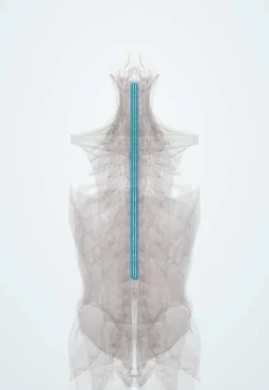
Posterior spinal arteries !
The posterior spinal arteries are two small arteries down the back of the spinal cord; they feed the part that carries position sense and fine touch.
Opens in the Durgical app, no sign-up needed.
What it is
Two small arteries running down the back of the spinal cord.
What it does
Supply the back third of the cord, which carries position sense and fine touch.
Key facts
- Origin
- Vertebral artery or posterior inferior cerebellar artery (PICA), one on each side
- Branches
- Paired longitudinal channels along the posterolateral sulci; reinforced by posterior segmental medullary arteries; pial plexus
- Supplies
- Posterior third of the cord: dorsal columns and posterior horns
Relations
- Course
- From the vertebral artery or the posterior inferior cerebellar artery beside the medulla, each descends along a posterolateral sulcus to the conus medullaris, joined at intervals by posterior segmental medullary arteries
- Medial
- The dorsal columns (fasciculus gracilis and fasciculus cuneatus), between the two arteries
- Lateral
- Dorsal nerve rootlets entering the cord
- Superficial
- Pia mater and, outside it, the subarachnoid space, the arachnoid and the dura
Examination
- Romberg test: steady with the eyes open but swaying or falling with them closed points to loss of joint position sense (dorsal columns)
- Vibration with a tuning fork and joint position at the big toe: loss below a level suggests dorsal column damage
On imaging
MRI shows bright T2 signal in the back of the cord; vitamin B12 deficiency (subacute combined degeneration) and other dorsal column disorders can look similar and are part of the differential.
Clinical notes
- A pial arterial network (vasocorona) around the cord links the posterior and anterior systems, which partly protects against a single blocked vessel
- Posterior spinal artery infarcts can be one-sided, so the sensory loss may be limited to one side of the body
- Dorsal column loss from vitamin B12 deficiency or tabes dorsalis comes on slowly; a sudden onset with restricted diffusion on MRI points instead to a posterior spinal artery infarct
Test yourself
Think of the answer, then tap the question to see it.
From which vessels do the posterior spinal arteries arise?
From the vertebral artery or the posterior inferior cerebellar artery (PICA), one on each side.
What part of the cord do the posterior spinal arteries supply?
The posterior third: the dorsal columns and the posterior horns.
What does a posterior spinal artery infarct cause, and why is it rare?
Loss of vibration and joint position sense below the level (sensory ataxia) with movement spared; rare because the paired arteries form a rich network.
Which bedside test points to dorsal column loss from a posterior spinal artery infarct?
The Romberg test: the person is steady with eyes open but sways or falls with them closed, from lost joint position sense.
Educational information, not medical advice. For any symptom, see a health professional.
Its names
- Official Latin (TA2)
- arteriae spinales posteriores TA2 4527
- Official Spanish (DeCS)
- Arterias espinales posteriores
- English (TA2)
- Posterior spinal arteries
- Also called
- arteriae spinales dorsales
Related
Sources: Terminologia Anatomica (TA2); Neuroanatomy, Spinal Cord; Tratado de anatomía humana (9.ª ed.), tomo II, Libro V Sistema nervioso central, sección 1: Médula espinal.
Spotted a mistake?We are always improving for the community. Tell us what is wrong and our team will review it.Report a mistake
Representative model, for educational purposes only.