Cardiovascular system · Abdomen
Appendicular artery !
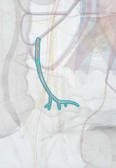
The appendicular artery is the only artery of the appendix, a branch of the ileocolic artery running in the mesoappendix to its tip.
Opens in the Durgical app, no sign-up needed.
What it is
A small artery in the mesoappendix, from the ileocolic artery.
What it does
The appendix's only blood supply; tied or sealed in an appendectomy.
Key facts
- Origin
- Ileocolic artery (often its posterior cecal or ileal branch), passing behind terminal ileum
- Branches
- Runs in free edge of mesoappendix giving small branches to the appendix (varies: accessory appendicular artery)
- Supplies
- Appendix; an end artery, so thrombosis leads to gangrene and perforation
Relations
- Course
- Leaves the ileocolic artery (or one of its cecal or ileal branches), passes behind the terminal ileum and enters the mesoappendix, running along its free edge toward the tip
- In front
- Terminal ileum, which crosses in front of it near its origin
- Lateral
- Cecum, where the base of the appendix lies at the meeting point of the taeniae coli
How to find it
The base of the appendix projects near McBurney point, one third of the way from the anterior superior iliac spine to the umbilicus. At surgery the taeniae coli are followed down to the base and the mesoappendix is spread out to show the artery near its free edge.
On imaging
Not seen on its own on routine imaging. It runs in the mesoappendix, so the appendix and the ileocolic vessels are the landmarks. In appendicitis, color Doppler can show more flow in the inflamed wall, while loss of flow suggests gangrene; CT shows a thick appendix with surrounding fat stranding. Ultrasound comes first in children and in pregnancy (MRI if unclear), CT in most adults.
Common problems
- Mesenteric ischemia: a clot or narrowing starves the gut; severe pain 'out of proportion' to the examination. An emergency.
How it is diagnosed
CT angiography.
Treatment
- Restore flow (catheter or surgery) and remove dead bowel.
Clinical notes
- In laparoscopic appendectomy the artery is sealed, clipped or tied within the mesoappendix; a poorly secured artery is a cause of early bleeding after surgery
- In a retrocecal appendix the mesoappendix is short and the artery lies behind the cecum, which makes it harder to expose
Anatomical variants
- Accessory appendicular artery from the posterior cecal artery
- The base of the appendix is constant on the cecum but the tip varies (retrocecal, subcecal, pelvic, in front of or behind the ileum), and the length and route of the artery change with it
Related conditions
Test yourself
Think of the answer, then tap the question to see it.
Where does the appendicular artery arise and run?
From the ileocolic artery (often its posterior cecal or ileal branch); it passes behind the terminal ileum and runs in the free edge of the mesoappendix.
Why does thrombosis of the appendicular artery lead to gangrene?
It is an end artery and the only blood supply of the appendix, so thrombosis causes gangrene and perforation.
What can cause bleeding after appendectomy even when the appendicular artery has been tied?
An accessory appendicular artery from the posterior cecal artery.
Educational information, not medical advice. For any symptom, see a health professional.
Its names
- Official Latin (TA2)
- arteria appendicularis TA2 4261
- Official Spanish (DeCS)
- Arteria apendicular
- English (TA2)
- Appendicular artery
Related
Sources: Terminologia Anatomica (TA2); Anatomy, Abdomen and Pelvis: Appendix.
Spotted a mistake?We are always improving for the community. Tell us what is wrong and our team will review it.Report a mistake
Representative model, for educational purposes only.