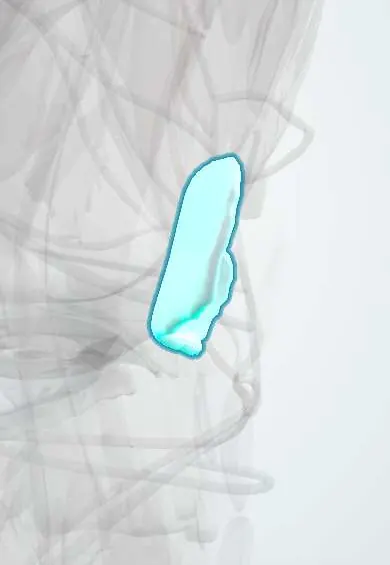
Patellar cartilage !
The patellar cartilage is the thick, smooth layer on the back of the kneecap; it glides on the femur and is heavily loaded on stairs.
Official name: Articular cartilage of patella
Opens in the Durgical app, no sign-up needed.
What it is
Thick articular cartilage on the back of the kneecap, gliding on the femur's groove.
What it does
Lets the joint glide with very little friction and spreads load across the bone.
Key facts
- Where
- Posterior articular surface of patella
- Function
- Hyaline articular cartilage; low-friction gliding; spreads load; thickest articular cartilage in the body; avascular, nourished by synovial fluid
Relations
- Behind
- Trochlear cartilage of the femur; in deep flexion the odd facet meets the medial femoral condyle
- Above
- Suprapatellar pouch
- Below
- Non-articular apex and the infrapatellar fat pad
- Deep
- Subchondral bone of the patella
How to find it
With the knee straight and relaxed, the patella is pushed to one side and the undersurface of its edge is pressed; tenderness there points to the facet cartilage.
Examination
- Patellar grind (Clarke) test: pain when the quadriceps contracts against a patella held down; common in normal knees too, so of low specificity
On imaging
Axial (skyline) view for patellofemoral joint-space narrowing and lateral tilt; MRI for fissures, defects and the bone-bruise pattern after a dislocation.
Common problems
- Softening and wear behind the kneecap (chondromalacia), often with pain on stairs or squatting.
- Osteoarthritis.
How it is diagnosed
Symptoms and exam; X-rays show joint-space narrowing in arthritis; MRI shows cartilage defects.
Treatment
- Physical therapy, weight management, activity changes; medicines or injections for pain.
- Surgery for focal defects: microfracture, cell-based repair, or osteochondral grafts (including osteochondral allograft from a donor).
- Knee replacement for advanced arthritis.
Clinical notes
- Excessive lateral pressure syndrome: a tight lateral retinaculum tilts the patella and overloads the lateral facet
- Osteochondral fragments after dislocation may be fixed back if large, or removed if small
- Chondromalacia patellae describes softening seen at surgery and does not always match the pain
Anatomical variants
- Bipartite patella: an accessory ossicle, usually at the upper outer corner, joined to the main bone by fibrocartilage; it is often bilateral and has smooth, rounded edges, unlike a fracture
Development
The patella forms as a sesamoid bone within the quadriceps tendon; it is cartilaginous at birth and ossifies in early childhood from several foci that merge, and an unfused focus explains the bipartite patella.
Related conditions
Test yourself
Think of the answer, then tap the question to see it.
Which activities load the patellar cartilage most, and why?
Squatting, kneeling and stairs: patellofemoral load rises with knee flexion and quadriceps force.
Which osteochondral injury follows a lateral patellar dislocation?
The medial patellar facet strikes the lateral femoral condyle, causing osteochondral fractures and loose bodies.
What is the odd facet of the patella?
The far medial edge of the patellar cartilage, which touches the medial femoral condyle only in deep flexion.
Educational information, not medical advice. For any symptom, see a health professional.
Its names
- Official Latin (TA2)
- cartilago articularis TA2 382
- Official Spanish (DeCS)
- Cartílago articular de la rótula DeCS D002358
- English (TA2)
- Articular cartilage of patella
Related
Sources: Terminologia Anatomica (TA2); DeCS; Anatomy, Bony Pelvis and Lower Limb, Knee; 9.6 Anatomy of Selected Synovial Joints (OpenStax (Rice University), 2022) · adapted, CC BY 4.0.
Spotted a mistake?We are always improving for the community. Tell us what is wrong and our team will review it.Report a mistake
Representative model, for educational purposes only.