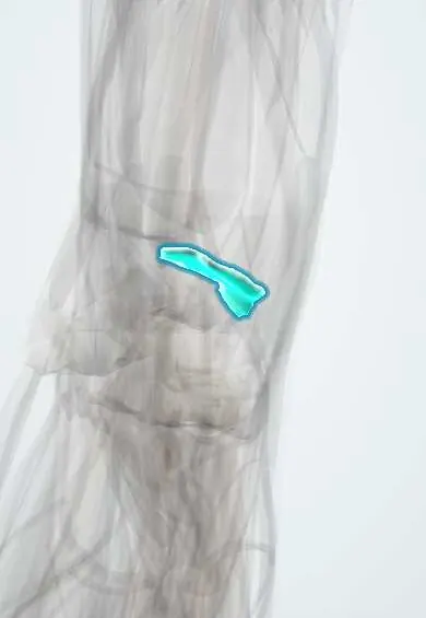
Wrist cartilage (radius) !
The wrist cartilage of the radius covers the end of the radius where the wrist bones sit; a fracture that leaves a step here can lead to arthritis.
Official name: Articular cartilage of the distal radius
Opens in the Durgical app, no sign-up needed.
What it is
The smooth surface at the end of the radius that meets the scaphoid and lunate.
What it does
Lets the wrist bend smoothly; a fracture that steps this surface can lead to arthritis.
Key facts
- Where
- Scaphoid and lunate fossae of the distal radius; ulnar notch
- Function
- Radiocarpal joint surface
Relations
- Below
- Scaphoid and lunate, across the radiocarpal joint
- Medial
- TFCC, whose articular disc attaches to the edge of the sigmoid notch, and the ulnar head turning in the notch
- In front
- Palmar wrist ligaments (radioscaphocapitate, long and short radiolunate), then pronator quadratus and the flexor tendons
- Behind
- Dorsal radiocarpal ligament and the extensor tendons; Lister's tubercle acts as a pulley for the EPL tendon
- Lateral
- Radial styloid with the brachioradialis tendon insertion and the first extensor compartment (APL and EPB tendons)
How to find it
Lister's tubercle is the bony bump on the back of the distal radius; the radiocarpal joint line lies about a fingertip's width beyond it. The 3–4 arthroscopy portal, between the EPL tendon and the finger extensors, enters the joint just there.
On imaging
PA and lateral X-rays measure inclination, height and tilt; CT shows step-offs, gaps and depression of the lunate fossa in intra-articular fractures; wrist arthroscopy inspects the cartilage directly.
Clinical notes
- Die-punch fracture: the lunate is driven into the lunate fossa, depressing that facet
- Barton fracture: a palmar or dorsal rim fracture with the carpus subluxing together with the fragment; chauffeur's fracture: an intra-articular fracture of the radial styloid
- A step left in the joint surface after a distal radius fracture predisposes to radiocarpal osteoarthritis, especially in younger, active people
Development
The distal end ossifies from a secondary centre that appears in early childhood and unites with the shaft in the late teens; this physis is the one most often injured in children, typically as a Salter–Harris type II fracture.
Related conditions
Test yourself
Think of the answer, then tap the question to see it.
Which facets form the articular surface of the distal radius?
The scaphoid fossa and the lunate fossa, separated by a low ridge, plus the sigmoid notch for the ulnar head.
In SLAC wrist, where does arthritis start and which joint is spared?
Between the radial styloid and the scaphoid; the radiolunate joint is typically spared.
What is a die-punch fracture of the distal radius?
The lunate is driven into the lunate fossa, depressing that facet.
Educational information, not medical advice. For any symptom, see a health professional.
Its names
- Official Latin (TA2)
- cartilago articularis facies articularis carpalis radii TA2 382
- Official Spanish (DeCS)
- Cartílago articular de la cara articular carpiana del radio
- English (TA2)
- Articular cartilage of the distal radius
Related
Sources: Terminologia Anatomica (TA2); Anatomy, Shoulder and Upper Limb, Hand Radiocarpal Joint; Anatomy, Shoulder and Upper Limb, Wrist Joint.
Spotted a mistake?We are always improving for the community. Tell us what is wrong and our team will review it.Report a mistake
Representative model, for educational purposes only.