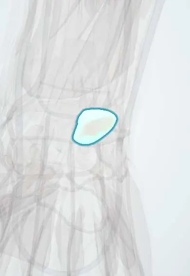
Lunate !
The lunate is the moon-shaped wrist bone in the centre of the first row; it sits between the radius and the capitate and can dislocate in falls.
Opens in the Durgical app, no sign-up needed.
What it is
The half-moon-shaped wrist bone in the centre, next to the radius.
What it does
One of the eight carpal bones that let the wrist bend and carry load to the forearm.
Key facts
- Joints with
- Radius, scaphoid, triquetrum, capitate (hamate); TFCC
- Landmarks
- Half-moon shape; most often dislocated carpal
Relations
- Above
- Lunate fossa of the distal radius laterally and the TFCC medially
- Below
- Head of the capitate in its concave distal surface; in many wrists also a small facet for the hamate
- Lateral
- Scaphoid, joined by the scapholunate ligament
- Medial
- Triquetrum, joined by the lunotriquetral ligament
- In front
- Palmar wrist ligaments; between them in front of the capitolunate joint lies a weak area (space of Poirier); further in front, the floor of the carpal tunnel with the median nerve and flexor tendons
- Behind
- Dorsal radiocarpal ligament and dorsal wrist capsule
How to find it
Felt on the back of the wrist just distal to Lister's tubercle, in line with the third metacarpal; it stands out when the wrist is flexed. Tenderness here is typical of Kienböck disease and of scapholunate injuries.
On imaging
Lateral view: radius, lunate and capitate should be collinear; dorsal tilt of the lunate is DISI and palmar tilt is VISI. PA view: three smooth carpal arcs (Gilula arcs) along the proximal row's upper and lower surfaces and the proximal capitate and hamate; a broken arc or a triangular lunate suggests dislocation. MRI shows early Kienböck changes before X-rays do.
Common problems
- Kienböck disease: the lunate slowly loses its blood supply and collapses.
- Dislocation in a severe wrist injury (perilunate dislocation), an emergency.
- Drifts out of line when the scapholunate ligament tears.
How it is diagnosed
X-rays, then MRI to see the blood supply. A dislocation is seen on the side X-ray.
Treatment
- Splint and activity change in early Kienböck disease.
- Surgery to unload or revascularize the lunate in later stages.
- Urgent reduction and repair for a dislocation.
Clinical notes
- Perilunate injuries progress from the radial to the ulnar side (Mayfield stages): scapholunate tear, capitolunate dislocation, lunotriquetral tear and finally lunate dislocation
- Perilunate dislocations are often missed on the first X-rays because the PA view can look almost normal
- A lunotriquetral ligament tear lets the lunate tilt palmarward with the triquetrum (VISI) and causes ulnar-sided wrist pain
Anatomical variants
- Some lunates have a separate medial facet that articulates with the hamate (type II lunate), linked to arthritis at the tip of the hamate
- Lunotriquetral coalition, the most common carpal coalition, usually painless and found by chance
Development
Ossifies around age 3–4, the fourth carpal bone to appear.
Related conditions
Test yourself
Think of the answer, then tap the question to see it.
On a lateral wrist X-ray, how does lunate dislocation differ from perilunate dislocation?
In lunate dislocation the lunate tips palmarward (spilled teacup sign); in perilunate dislocation the capitate leaves the lunate's cup.
Which nerve can a lunate dislocated into the carpal tunnel compress?
The median nerve, causing acute carpal tunnel syndrome.
What is Kienböck disease, and which ulnar variance is it linked to?
Loss of blood supply and collapse of the lunate; it is associated with negative ulnar variance.
In what order do perilunate injuries progress (Mayfield stages)?
Radial to ulnar: scapholunate tear, capitolunate dislocation, lunotriquetral tear and finally lunate dislocation.
Educational information, not medical advice. For any symptom, see a health professional.
Its names
- Official Latin (TA2)
- os lunatum TA2 1252
- Official Spanish (DeCS)
- Hueso semilunar
- English (TA2)
- Lunate
Related
- Dorsal intercarpal ligament
- Dorsal radiocarpal ligament
- Extensor tendons
- FDP tendons
- Lunotriquetral ligament
- Midcarpal ligaments
- Scapholunate ligament
- TFCC
Sources: Terminologia Anatomica (TA2); Anatomy, Shoulder and Upper Limb, Hand Carpal Bones.
Spotted a mistake?We are always improving for the community. Tell us what is wrong and our team will review it.Report a mistake
Representative model, for educational purposes only.