Joints and ligaments · Upper limb
TFCC !
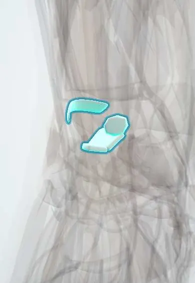
The triangular fibrocartilage complex (TFCC) cushions the little-finger side of the wrist and holds the radius and ulna together as the forearm turns.
Official name: Triangular fibrocartilage complex
Opens in the Durgical app, no sign-up needed.
What it is
A cushion of cartilage and ligaments between the end of the ulna and the wrist, on the little-finger side.
What it does
Cushions the ulnar side of the wrist and holds the radius and ulna together as the forearm turns.
Key facts
- Attaches
- Articular disc from the ulnar notch of the radius to the ulnar fovea and styloid, with dorsal and palmar radioulnar ligaments
- Function
- Stabilises the distal radioulnar joint; cushions the ulnar side of the wrist (loaded in ulnar deviation)
Relations
- Above
- Head of the ulna and the DRUJ cavity, which the disc seals off from the radiocarpal joint
- Below
- Lunate and triquetrum, which glide on its distal surface; the ulnocarpal ligaments blend with its palmar edge
- Lateral
- Ulnar (sigmoid) notch of the radius, where the disc and both radioulnar ligaments arise
- Medial
- Ulnar styloid and fovea; the meniscus homologue and the ulnar collateral ligament on the far ulnar side
- Behind
- Extensor carpi ulnaris (ECU) tendon in its subsheath in the sixth dorsal compartment, which is counted as part of the complex
How to find it
With the forearm in neutral rotation, the soft spot bounded by the ulnar styloid, the flexor carpi ulnaris tendon, the pisiform and the palmar ulnar head lies over the foveal attachment. The disc itself sits just distal to the ulnar head, on the little-finger side of the wrist.
Examination
- Fovea sign: pressure in the soft spot between the ulnar styloid and flexor carpi ulnaris reproduces the pain; points to a foveal TFCC detachment or an ulnotriquetral ligament tear
- TFCC load (ulnocarpal stress) test: axial load with the wrist in ulnar deviation while the forearm is rotated; pain, clicking or crepitus suggests a TFCC tear or ulnar impaction
- DRUJ ballottement (piano-key test): the radius is held and the ulnar head pushed dorsally and palmarly, compared with the other side; extra glide means DRUJ instability, usually from a foveal tear
On imaging
A PA view in neutral rotation (shoulder abducted, elbow at 90°) measures ulnar variance. MRI, or better MR arthrography, shows a tear and whether it is central or peripheral; contrast passing from the radiocarpal joint into the DRUJ marks a perforation. Arthroscopy remains the reference standard: a torn disc loses its normal springy tension when probed.
Common problems
- Tear from a fall on an outstretched hand or from twisting; pain on the little-finger side of the wrist.
- Wear with age, especially when the ulna is a little longer than the radius (ulnar impaction).
- Clicking and weakness when turning a doorknob or lifting a pan.
How it is diagnosed
Tenderness in the soft spot next to the ulnar head and pain with twisting tests. X-rays check ulnar length; MRI or arthroscopy shows the tear.
Treatment
- Splint, activity changes and sometimes an injection.
- Arthroscopic trimming or repair of the tear.
- Ulnar shortening surgery when the ulna is too long.
Clinical notes
- Distal radius fractures with a fracture at the base of the ulnar styloid can take the foveal attachment with them and leave the DRUJ unstable
- Galeazzi fracture-dislocation: a radial shaft fracture with disruption of the DRUJ, in which the TFCC is torn
- Ulnar-sided wrist pain has a crowded differential around it: ECU tendinitis or subluxation, lunotriquetral ligament tear, pisotriquetral arthritis, hook of hamate fracture
- Ulnar shortening osteotomy unloads the TFCC when ulnar impaction comes with positive variance
Anatomical variants
- Central perforations of the disc become common with age and are often degenerative and painless, so an MRI finding needs clinical correlation
- Ulnar variance (length of the ulnar head relative to the radius) differs between people; the disc tends to be thicker with negative variance and thinner with positive variance
Related conditions
Test yourself
Think of the answer, then tap the question to see it.
What is the main stabiliser of the distal radioulnar joint?
The TFCC, through its dorsal and palmar radioulnar ligaments anchored deep in the ulnar fovea.
Why can peripheral TFCC tears heal while central tears usually cannot?
Only the outer rim of the disc has a blood supply; central tears are usually debrided.
How does the Palmer classification divide TFCC tears?
Class 1 traumatic (1A central, 1B ulnar, 1C distal, 1D radial) and class 2 degenerative, linked to ulnar-positive variance.
What does the fovea sign point to?
A foveal TFCC detachment or an ulnotriquetral ligament tear: pressure between the ulnar styloid and flexor carpi ulnaris reproduces the pain.
Which operation unloads the TFCC in ulnar impaction with positive variance?
Ulnar shortening osteotomy.
Educational information, not medical advice. For any symptom, see a health professional.
Its names
- Official Latin (TA2)
- discus articulationis radioulnaris distalis TA2 1782
- Official Spanish (DeCS)
- Disco articular de la articulación radioulnar distal; ligamentos radioulnares dorsal y palmar
- English (TA2)
- Triangular fibrocartilage complex
- Also called
- Articular disc of the distal radio-ulnar joint (its main part)
Related
Sources: Terminologia Anatomica (TA2); Anatomy, Shoulder and Upper Limb, Forearm Triangular Fibrocartilage Complex; Anatomy, Shoulder and Upper Limb, Distal Radio-Ulnar Joint.
Spotted a mistake?We are always improving for the community. Tell us what is wrong and our team will review it.Report a mistake
Representative model, for educational purposes only.