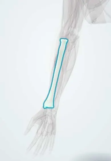
Distal radius fracture (Colles) !
A distal radius fracture is a break of the forearm bone just above the wrist, often from a fall on the hand; in the Colles type the wrist looks like a fork.
Updated 2026-10-29
Structures involved
Tap one to see what it is and open it in 3D.
In histology
Why it happens
What it is
A fracture of the distal radius, typically within a few centimetres of the wrist. In the Colles type, the distal fragment is displaced and angled dorsally, giving the classic «dinner fork» deformity.
Who gets it
One of the most common fractures in adults. It peaks in children and young people after high-energy trauma, and in older women with osteoporosis after simple falls; it is often the first sign of osteoporosis.
Causes
- Fall on an outstretched hand with the wrist extended (Colles)
- Fall on a flexed wrist (Smith fracture, volar displacement)
- High-energy trauma in younger patients
Risk factors
- Osteoporosis
- Female sex and age over about 50
- Falls and poor balance
- Contact sports and road traffic accidents in the young
Pathophysiology
Landing on the extended wrist loads the metaphysis of the distal radius, which fails in tension on the palmar side and is compressed dorsally. The distal fragment tilts and shifts dorsally and radially and the radius shortens. In osteoporotic bone the dorsal cortex comminutes, which makes the reduction harder to hold.
Classification
- Extra-articular or intra-articular
- Colles (dorsal displacement), Smith (volar displacement), Barton (rim fracture-dislocation), chauffeur (radial styloid)
- Stable or unstable; formal systems such as AO/OTA and Frykman are used
Symptoms and signs
- Pain and swelling of the wrist after a fall
- Dinner-fork deformity
- Tenderness over the distal radius
- Reduced wrist and forearm movement
- Tingling in the median nerve fingers if the nerve is compressed
Diagnosis
- Clinical
- Inspection for deformity and skin breaks; examination of median nerve sensation, radial pulse and capillary refill; check the elbow, scaphoid and distal radioulnar joint.
- Tests
- In older adults after a low-energy fracture, bone density testing (DXA) and metabolic labs as part of osteoporosis assessment.
- Imaging
- Posteroanterior and lateral wrist X-rays assess radial height, inclination and dorsal tilt. CT for complex intra-articular fractures.
Differential diagnosis
- Scaphoid fracture
- Wrist sprain or scapholunate ligament injury
- Distal radioulnar joint injury
- Smith (reverse Colles) fracture
- Perilunate dislocation
Goal of treatment
Restore acceptable alignment and joint congruity, protect the median nerve and regain a pain-free, functional wrist; treat underlying osteoporosis.
Treatment
- Acute
- Closed reduction under regional or local anaesthesia (such as a haematoma block) for displaced fractures, with post-reduction X-ray.
- Conservative
- Immobilisation in a splint or cast for stable or well-reduced fractures, with repeat X-rays to detect loss of position.
- Surgical
- Open reduction and internal fixation, usually with a volar plate, or pinning or external fixation for unstable, intra-articular or re-displaced fractures.
- Rehabilitation
- Finger movement from the start and wrist physiotherapy after immobilisation to recover motion and grip.
- Medical
- Assessment and treatment of osteoporosis in older adults to prevent further fragility fractures.
Complications
- Malunion and loss of reduction
- Median nerve compression (acute carpal tunnel syndrome)
- Complex regional pain syndrome
- Extensor pollicis longus tendon rupture
- Stiffness and post-traumatic arthritis
- Distal radioulnar joint dysfunction
Prognosis
Most patients regain good function. Older, lower-demand patients often do well even with some residual deformity, while younger patients need closer restoration of anatomy to avoid later arthritis.
Prevention
- Fall prevention and home safety in older adults
- Osteoporosis screening and treatment
- Wrist guards in sports such as skating
Test yourself
Think of the answer, then tap the question to see it.
What is the typical mechanism and displacement of a Colles fracture?
A fall on an outstretched hand with the wrist extended; the distal radius fragment is displaced and angled dorsally (dinner-fork deformity).
How does a Smith fracture differ from a Colles fracture?
In a Smith fracture the distal fragment is displaced volarly, usually after a fall on a flexed wrist.
Which nerve is most at risk in a distal radius fracture?
The median nerve, which can develop acute carpal tunnel syndrome.
Which underlying condition should be considered in an older woman with a low-energy distal radius fracture?
Osteoporosis; the fracture is a fragility fracture and warrants bone health assessment.
Which tendon can rupture weeks after an undisplaced distal radius fracture?
The extensor pollicis longus, near Lister's tubercle.
Related conditions
- Carpal tunnel syndrome
- Scaphoid fracture
- Osteoporotic vertebral compression fracture
- Boxer fracture (fifth metacarpal neck)
- Elbow dislocation
Educational information, not medical advice. For any symptom, see a health professional.
Sources: Colles Fracture (StatPearls (NCBI Bookshelf), 2023).
Spotted a mistake?We are always improving for the community. Tell us what is wrong and our team will review it.Report a mistake
Representative model, for educational purposes only.