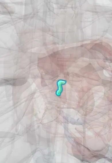
Bundle of His !
The bundle of His is a short band of conducting fibres that carries each heartbeat signal from the atrioventricular node into the ventricles.
Official name: Atrioventricular bundle
Opens in the Durgical app, no sign-up needed.
What it is
A short cable of conducting fibres carrying the signal from the AV node through the heart's fibrous skeleton into the septum.
What it does
Passes the beat to the bundle branches.
Key facts
- Where
- From AV node through right fibrous trigone (central fibrous body) along lower edge of membranous septum
- Function
- Only electrical link through fibrous skeleton; divides at top of muscular septum into right and left bundle branches
Relations
- Course
- From the AV node it pierces the central fibrous body (right fibrous trigone), runs forward under the membranous septum along the top of the muscular septum and divides into the right and left bundle branches
- Above
- Membranous interventricular septum; on the left side, the area below the commissure between the right and non-coronary cusps of the aortic valve
How to find it
In the electrophysiology laboratory a catheter laid across the tricuspid valve at the top of the septum, just below the anteroseptal commissure, records the His spike; in a dissection the bundle is followed from the apex of the triangle of Koch to the crest of the muscular septum.
Examination
- His bundle electrogram in an electrophysiology study: the His spike separates the AH interval (AV node) from the HV interval (His–Purkinje system); a prolonged HV interval places the block below the node
On imaging
Not seen on imaging; it is recorded as the His deflection in an electrophysiology study. Its path is inferred from the membranous septum just below the aortic valve, seen on echocardiography and cardiac CT. Before transcatheter aortic valve implantation, CT assesses this septum, since a short one is linked to a higher risk of heart block afterwards.
Common problems
- Heart block: the signal is slowed or blocked, causing a slow pulse and fainting.
- Re-entry circuits around it cause sudden fast heartbeats (SVT).
How it is diagnosed
ECG (PR interval), Holter monitor, electrophysiology study.
Treatment
- Pacemaker for heart block; catheter ablation for SVT.
Clinical notes
- Heart block after ventricular septal defect closure, aortic valve replacement or transcatheter aortic valve implantation, because the bundle runs just beneath the aortic annulus
- His bundle pacing places the lead on the bundle to use the heart's own conduction system and produce a narrow QRS
- A junctional escape rhythm from the AV junction and His region has a narrow QRS at about 40–60 beats per minute
- Lenègre–Lev disease: age-related fibrosis and calcification of the conduction system and the fibrous skeleton around it, a common cause of heart block in older adults
Related conditions
Test yourself
Think of the answer, then tap the question to see it.
What makes the bundle of His unique in the atrioventricular junction?
It is normally the only muscular electrical link between atria and ventricles, piercing the central fibrous body (right fibrous trigone).
Why is the bundle of His at risk in septal defect repair and aortic valve procedures?
It runs along the lower edge of the membranous septum on the crest of the muscular septum, just below the aortic valve.
In an electrophysiology study, what does a prolonged HV interval indicate?
A block below the AV node, in the His–Purkinje system; the His spike separates the AH (AV node) from the HV interval.
What are the QRS width and rate of a junctional escape rhythm?
A narrow QRS at about 40–60 beats per minute, from the AV junction and the His region.
Educational information, not medical advice. For any symptom, see a health professional.
Its names
- Official Latin (TA2)
- fasciculus atrioventricularis TA2 3955
- Official Spanish (DeCS)
- Fascículo atrioventricular DeCS D002036
- English (TA2)
- Atrioventricular bundle
- Also called
- truncus fasciculi atrioventricularis · Kent's bundle
Sources: Terminologia Anatomica (TA2); DeCS; Physiology, AV Junction; Anatomy, Thorax, Heart.
Spotted a mistake?We are always improving for the community. Tell us what is wrong and our team will review it.Report a mistake
Representative model, for educational purposes only.