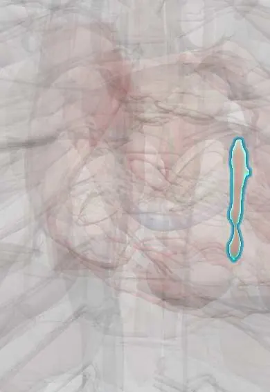
Interventricular septum !
The interventricular septum is the wall between the two lower chambers of the heart; it keeps oxygen-rich and oxygen-poor blood apart.
Opens in the Durgical app, no sign-up needed.
What it is
The thick muscular wall between the two ventricles, with a thin membranous part near the top.
What it does
Separates oxygen-rich from oxygen-poor blood and carries the bundle branches of the conduction system.
Key facts
- Where
- Between ventricles; thick muscular part and small membranous part just below aortic valve
- Function
- Separates ventricles; carries bundle branches; supplied by LAD septals (anterior 2/3) and PDA (posterior 1/3); membranous part = commonest VSD site
Relations
- In front
- Anterior interventricular groove with the LAD (anterior descending artery), which sends septal perforating branches into it
- Behind
- Posterior interventricular groove with the posterior descending artery (PDA) and the middle cardiac vein
- Above
- Aortic valve above the membranous part; the tricuspid septal leaflet attaches across the membranous septum on its right side
- Contents
- The left bundle branch fans out under the left ventricular endocardium; the right bundle branch runs down to the moderator band
How to find it
It cannot be felt. A ventricular septal defect gives a loud, harsh holosystolic murmur at the left lower sternal border, often with a thrill; small defects are typically the loudest.
On imaging
Echocardiography: parasternal long and short axis and apical four-chamber views measure its thickness and show defects with colour Doppler. Septal motion becomes paradoxical with left bundle branch block or after heart surgery. Cardiac MRI shows septal scar.
Common problems
- A hole from birth (ventricular septal defect).
- Can rupture after a large heart attack (rare).
How it is diagnosed
ECG, blood tests (troponin) for a heart attack; echocardiogram for ejection fraction and wall movement; cardiac MRI.
Treatment
- Opening the blocked artery fast in a heart attack (angioplasty and stent).
- Heart-failure medicines, and devices such as a defibrillator or resynchronization pacemaker.
- Bypass surgery when several arteries are narrowed.
Clinical notes
- Many small muscular defects close by themselves in childhood; large defects cause heart failure in infancy and, left alone, Eisenmenger syndrome
- Post-infarct rupture is apical after anterior infarcts and basal-inferior after inferior infarcts
- Hypertrophic cardiomyopathy thickens the septum asymmetrically and can narrow the left ventricular outflow tract
- Closing a perimembranous defect risks the bundle of His at its lower rim, causing heart block
- The septal tricuspid leaflet hinges below the top of the membranous septum, so its upper (atrioventricular) part lies between the left ventricle and the right atrium; a defect there shunts left ventricle to right atrium (Gerbode defect)
Development
The muscular part grows up from the floor of the primitive ventricle; the membranous part closes last, from the endocardial cushions and the outflow ridges, which is why defects cluster there.
Related conditions
- Hypertrophic cardiomyopathy
- Mechanical complications of myocardial infarction
- Eisenmenger syndrome
- Down syndrome (trisomy 21)
- Rubella and congenital rubella syndrome
- Trisomy 18 (Edwards) and trisomy 13 (Patau)
- Hypertensive heart disease
- Ventricular septal defect
- Tetralogy of Fallot
- Myocardial Infarction (Heart Attack)
Test yourself
Think of the answer, then tap the question to see it.
Where is the commonest ventricular septal defect located?
In the membranous part of the interventricular septum (perimembranous), just below the aortic valve, which closes last in development.
What murmur does a ventricular septal defect produce?
A loud, harsh holosystolic murmur at the left lower sternal border, often with a thrill; small defects are typically the loudest.
How does post-infarction rupture of the interventricular septum present?
A new harsh holosystolic murmur and sudden heart failure, usually within the first days after the infarct.
Which arteries supply the interventricular septum?
Septal perforators of the LAD supply the anterior 2/3; the posterior descending artery supplies the posterior 1/3.
Why does closing a perimembranous ventricular septal defect risk heart block?
The bundle of His runs along the lower edge of the membranous septum, at the lower rim of the defect.
Educational information, not medical advice. For any symptom, see a health professional.
Its names
- Official Latin (TA2)
- septum interventriculare TA2 3970
- Official Spanish (DeCS)
- Tabique interventricular DeCS D054088
- English (TA2)
- Interventricular septum
- Also called
- ventricular septum · septum ventriculorum
Related
Sources: Terminologia Anatomica (TA2); DeCS; Anatomy, Thorax, Heart; Anatomy, Thorax, Heart and Pericardial Cavity; Tratado de anatomía humana (9.ª ed.), tomo II, Libro IV Angiología, sección 1, capítulo 1: Corazón, artículos I–VI.
Spotted a mistake?We are always improving for the community. Tell us what is wrong and our team will review it.Report a mistake
Representative model, for educational purposes only.