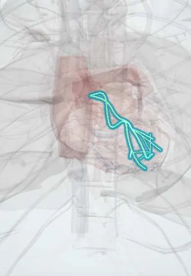
Bundle branches !
The bundle branches are the right and left paths of the heart's wiring in the septum; they spread the signal so both ventricles contract together.
Official name: Right bundle branch
Opens in the Durgical app, no sign-up needed.
What it is
The right and left branches of the conduction system running down the septum and spreading into the ventricle walls as Purkinje fibres.
What it does
Deliver the signal so both ventricles contract together, from the apex upward.
Key facts
- Where
- Right branch: right side of septum, via moderator band to anterior papillary muscle; left branch: left septal surface, anterior and posterior fascicles
- Function
- Carry impulse to Purkinje fibers so septum and apex contract first, then base; block widens QRS
Relations
- Course
- The right bundle branch runs down the right side of the septum, deep at first, then through the septomarginal trabecula (moderator band) to the anterior papillary muscle; the left bundle branch fans out under the left septal endocardium into anterior and posterior fascicles (some texts add a septal fascicle)
- Superficial
- Endocardium of the interventricular septum; from the branches, Purkinje fibers spread beneath the endocardium of both ventricles
- Above
- Aortic valve: the left bundle branch leaves the His bundle just below the commissure between the right and non-coronary cusps, so aortic valve surgery or transcatheter valve implantation often causes new left bundle branch block
How to find it
Not visible from outside. In the opened heart the bundle of His runs along the lower edge of the membranous septum; the right branch descends on the right face of the septum and crosses to the anterior papillary muscle inside the moderator band, while the left branch fans out under the endocardium of the left face. Sutures in this zone, as in VSD repair, can cause block.
On imaging
Not seen on imaging; their function is read on the ECG. Echocardiography shows the mechanical effect of left bundle branch block, an early inward flick of the septum (septal flash) and out-of-step left ventricular contraction, assessed before resynchronization. Cardiac MRI with late gadolinium looks for the scar behind it, such as an old infarct or Chagas disease.
Common problems
- Bundle branch block (seen on ECG): the ventricles contract out of step; a left block can worsen heart failure.
How it is diagnosed
ECG (PR interval), Holter monitor, electrophysiology study.
Treatment
- A resynchronization pacemaker (CRT) re-coordinates the ventricles in selected heart-failure patients.
Clinical notes
- Left bundle branch block hides ischemic ST changes on the ECG; Sgarbossa criteria are used to look for infarction despite it
- Left bundle branch block makes the ventricles contract out of step; in heart failure, cardiac resynchronization adds a left ventricular lead through the coronary sinus to restore synchrony
- The right bundle branch is thin and close to the endocardium for part of its course, so a catheter in the right ventricle can bruise it and cause temporary right bundle branch block
- Right bundle branch block with left anterior fascicular block (bifascicular block) or alternating bundle branch block signals disease of the whole His–Purkinje system and a risk of complete heart block
- Septal perforators of the LAD supply most of the bundle branches, so an anterior MI can cause new bundle branch block, a sign of a large infarct
Memory aid
WiLLiaM MaRRoW: in LBBB the QRS looks like a W in V1 and an M in V6; in RBBB, an M in V1 and a W in V6.
Related conditions
- Ventricular tachycardia
- Long QT syndrome and torsades de pointes
- Brugada syndrome
- Chagas cardiomyopathy
- Atrioventricular (heart) block
Test yourself
Think of the answer, then tap the question to see it.
What are the ECG criteria for right bundle branch block?
QRS 0.12 s or wider, rSR′ in V1–V2 and a broad S wave in I and V6.
What are the ECG criteria for left bundle branch block?
QRS 0.12 s or wider, broad notched R waves in I, aVL and V5–V6, no septal q waves and a deep S wave in V1.
Why does the left anterior fascicle block more often than the posterior, and what axis results?
It is long and thin, while the posterior fascicle is broad with a double blood supply; anterior fascicular block gives left axis deviation.
Which conduction pattern is classic in Chagas disease?
Right bundle branch block with left anterior fascicular block.
Why is a new bundle branch block in an anterior myocardial infarction a sign of a large infarct?
Septal perforators of the LAD supply most of the bundle branches.
Educational information, not medical advice. For any symptom, see a health professional.
Its names
- Official Latin (TA2)
- crus dextrum fasciculi atrioventricularis; crus sinistrum fasciculi atrioventricularis; rami subendocardiales TA2 3956
- Official Spanish (DeCS)
- Ramas derecha e izquierda del fascículo auriculoventricular DeCS D011690
- English (TA2)
- Right bundle branch
- Also called
- right branch of atrioventricular bundle · left branch of atrioventricular bundle
Related
Sources: Terminologia Anatomica (TA2); DeCS; Physiology, AV Junction; Anatomy, Thorax, Heart; 19.2 Cardiac Muscle and Electrical Activity (OpenStax (Rice University), 2022) · adapted, CC BY 4.0; Tratado de anatomía humana (9.ª ed.), tomo II, Libro IV Angiología, sección 1, cap. I Corazón (arts. VI–IX) y cap. II Pericardio.
Spotted a mistake?We are always improving for the community. Tell us what is wrong and our team will review it.Report a mistake
Representative model, for educational purposes only.