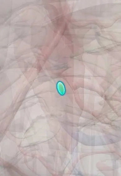
Atrioventricular (AV) node !
The atrioventricular node, low in the right atrium, is the electrical gateway from atria to ventricles; it briefly delays each beat so they fill.
Official name: Atrioventricular node
Opens in the Durgical app, no sign-up needed.
What it is
A small node of conducting tissue low in the wall between the atria, just above the ventricles.
What it does
The only electrical gateway from the atria to the ventricles: it delays the signal for a split second so the atria finish filling the ventricles first.
Key facts
- Where
- Interatrial septum in triangle of Koch, near coronary sinus opening; AV nodal artery usually from RCA (~80%)
- Function
- Delays impulse (about 0.1 s) so atria empty before ventricles contract; only normal atria-to-ventricle pathway
Relations
- In front
- Bundle of His, which it becomes as it enters the central fibrous body below the membranous septum
- Behind
- Opening of the coronary sinus at the base of the triangle of Koch; the slow-pathway region lies between it and the tricuspid annulus
- Above
- Tendon of Todaro, a fibrous cord running from the valve of the inferior vena cava to the central fibrous body
- Below
- Attachment of the septal leaflet of the tricuspid valve
How to find it
Found from inside the right atrium using the triangle of Koch: the node lies near the apex of the triangle, just below the membranous septum, where the tendon of Todaro and the septal tricuspid leaflet hinge converge. Electrophysiologists use the same landmarks on fluoroscopy and electroanatomic maps.
Examination
- Vagal maneuvers (Valsalva, carotid sinus massage) slow conduction through the node: they can stop a re-entrant tachycardia that uses the node, or slow the ventricular rate enough to reveal atrial flutter waves
- Electrophysiology study: the AH interval measures conduction through the node; a long AH places a block at the node rather than below it
On imaging
Not seen on imaging; AV conduction is judged on the ECG from the PR interval. The node is placed by the landmarks of the triangle of Koch (coronary sinus ostium, septal tricuspid leaflet, membranous septum), shown on echocardiography and cardiac CT and used in electroanatomic mapping. Coronary CT angiography can show the AV nodal artery at the crux.
Common problems
- Heart block: the signal is slowed or blocked, causing a slow pulse and fainting.
- Re-entry circuits around it cause sudden fast heartbeats (SVT).
How it is diagnosed
ECG (PR interval), Holter monitor, electrophysiology study.
Treatment
- Pacemaker for heart block; catheter ablation for SVT.
Clinical notes
- AV nodal re-entrant tachycardia, the commonest regular narrow-QRS tachycardia, uses a fast and a slow pathway; ablation targets the slow pathway between the coronary sinus ostium and the tricuspid annulus, away from the compact node
- In an inferior MI, ischemia of the AV nodal artery causes first-degree or Wenckebach block, usually with a narrow QRS and often temporary
- Operations near the triangle of Koch (tricuspid or mitral valve repair, septal defect closure) carry a risk of complete heart block
- Pre-excitation: an accessory pathway bypasses the node's delay, giving a short PR interval and a delta wave (Wolff–Parkinson–White pattern)
Anatomical variants
- The AV nodal artery arises from the artery that crosses the crux: the right coronary in about 80% of hearts (right dominance), the circumflex in most of the rest
Development
Forms from specialised muscle near where the sinus venosus meets the atrioventricular canal, at the back of the developing septum; this is why it ends up near the coronary sinus opening.
Related conditions
- Supraventricular tachycardia (AVNRT, AVRT)
- Wolff-Parkinson-White syndrome
- Digoxin toxicity
- Beta-blocker and calcium-channel-blocker overdose
- Atrial fibrillation
- Atrioventricular (heart) block
Test yourself
Think of the answer, then tap the question to see it.
Which landmarks bound the triangle of Koch, where the AV node lies?
The tendon of Todaro, the hinge of the septal tricuspid leaflet and, at its base, the coronary sinus ostium; the node lies near the apex.
Which part of the PR interval is due to the AV node?
Most of it: its slow conduction delays the impulse so the atria empty first (normal PR 0.12–0.20 s).
Which heart blocks are usually at the AV node, and which lie below it?
First-degree and Mobitz I (Wenckebach) are usually at the node; Mobitz II and complete block with a wide QRS usually lie in the His–Purkinje system.
In AV nodal re-entrant tachycardia, what does ablation target?
The slow pathway, between the coronary sinus ostium and the tricuspid annulus, away from the compact AV node.
How do vagal maneuvers act on the AV node?
They slow conduction through it: they can stop a re-entrant tachycardia using the node, or slow the rate enough to reveal atrial flutter waves.
Educational information, not medical advice. For any symptom, see a health professional.
Its names
- Official Latin (TA2)
- nodus atrioventricularis TA2 3954
- Official Spanish (DeCS)
- Nodo atrioventricular DeCS D001283
- English (TA2)
- Atrioventricular node
- Also called
- AV node · nodus fasciculi atrioventricularis · node of Tawara · aschoff-Tawara node
Related
Sources: Terminologia Anatomica (TA2); DeCS; Physiology, AV Junction; Anatomy, Thorax, Heart.
Spotted a mistake?We are always improving for the community. Tell us what is wrong and our team will review it.Report a mistake
Representative model, for educational purposes only.