Nervous system · Head and neck
Occipital lobe !
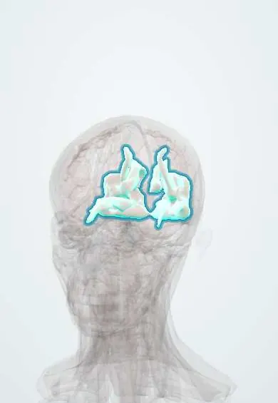
The occipital lobe is the back part of each half of the brain, at the back of the head; it processes vision.
Official name: Superior occipital gyri
Opens in the Durgical app, no sign-up needed.
What it is
The back of each hemisphere.
What it does
Processes vision.
Key facts
- Where
- Back of the hemisphere behind the parieto-occipital sulcus
- Blood supply
- PCA
- Function
- Vision
Relations
- In front
- Parietal lobe (parieto-occipital sulcus on the medial surface) and, below, the temporal lobe
- Behind
- Occipital bone; the transverse sinus runs along the tentorial attachment at its lower edge
- Below
- Tentorium, with the cerebellum beneath it
- Medial
- Falx cerebri; on the medial surface the calcarine sulcus with the visual cortex, the cuneus above and the lingual gyrus below
- Deep
- Occipital horn of the lateral ventricles and the end of the optic radiation
How to find it
The parieto-occipital sulcus on the medial surface is its clear front border; the calcarine sulcus runs forward from the occipital pole and joins it in a Y. Laterally there is no dividing sulcus, only a line to the preoccipital notch. From outside, the lobe lies above the level of the inion (external occipital protuberance), which overlies the confluence of sinuses.
Examination
- Confrontation visual fields, each quadrant of each eye: a matching (homonymous) defect on the same side in both eyes places the lesion behind the optic chiasm
- Pupillary light reflex: preserved in cortical blindness; reduced when the eye, optic nerve or chiasm is the cause
On imaging
Diffusion-weighted MRI shows a posterior cerebral artery infarct along the medial occipital lobe. Posterior reversible encephalopathy syndrome (with severe high blood pressure, eclampsia or some immunosuppressants) shows vasogenic oedema on FLAIR, mainly in the parieto-occipital white matter and cortex of both sides.
Common problems
- Head injury and tumours here change personality, judgement and planning.
- Front-lobe dementia changes behaviour first.
How it is diagnosed
Neurological and memory tests; CT or MRI.
Treatment
- Surgery for tumours when possible.
- Rehabilitation and support.
Clinical notes
- Alexia without agraphia: a left posterior cerebral artery stroke that also takes the splenium of the corpus callosum; the person can write but cannot read what was written
- Visual migraine aura, a shimmering zigzag scotoma that spreads over minutes, is attributed to spreading depression across the occipital cortex
Related conditions
- MELAS (mitochondrial encephalomyopathy, lactic acidosis and stroke-like episodes)
- Dementia with Lewy bodies
- Sturge–Weber syndrome
- Hallucinogen and dissociative drug intoxication (LSD, PCP, ketamine)
- Epilepsy and seizures
Test yourself
Think of the answer, then tap the question to see it.
What visual field defect does a posterior cerebral artery stroke of the occipital lobe cause?
A contralateral homonymous hemianopia, often with macular sparing, as the occipital pole may also get middle cerebral artery branches.
Why are pupillary light reflexes normal in cortical blindness?
The reflex fibres leave the visual pathway before the cortex; some people with bilateral occipital damage deny being blind (Anton syndrome).
What are the two visual outflow streams from the occipital lobe?
Dorsal to the parietal lobe (where things are, motion) and ventral to the temporal lobe (what they are: objects, faces, colour).
What causes alexia without agraphia?
A left posterior cerebral artery stroke that also takes the splenium of the corpus callosum: the person can write but cannot read what was written.
Educational information, not medical advice. For any symptom, see a health professional.
Its names
- Official Latin (TA2)
- gyri occipitales superiores TA2 5481
- Official Spanish (DeCS)
- Lóbulo Occipital DeCS D009778
- English (TA2)
- Superior occipital gyri
Related
Sources: Terminologia Anatomica (TA2); DeCS; Neuroanatomy, Occipital Lobe.
Spotted a mistake?We are always improving for the community. Tell us what is wrong and our team will review it.Report a mistake
Representative model, for educational purposes only.