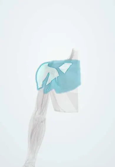
Shoulder fasciae !
The shoulder fasciae are sheets covering the deltoid and chest muscles; the cephalic vein passes through one of them, the clavipectoral fascia.
Official name: Deltoid, pectoral and clavipectoral fasciae
Opens in the Durgical app, no sign-up needed.
What it is
Thin sheets over the deltoid and chest muscles.
What it does
Cover the shoulder and chest muscles; the cephalic vein pierces the clavipectoral fascia.
Key facts
- Where
- Deltoid, pectoral and clavipectoral fasciae
- Function
- Cover the shoulder and chest muscles; the clavipectoral fascia is pierced by the cephalic vein, thoracoacromial artery and lateral pectoral nerve
Relations
- Superficial
- Pectoral fascia on the pectoralis major with the breast in front; deltoid fascia over the deltoid, attached to clavicle, acromion and scapular spine
- Deep
- Clavipectoral fascia deep to the pectoralis major, around the subclavius and pectoralis minor; behind it, the axillary artery, axillary vein and the cords of the brachial plexus
- Contents
- Structures piercing the clavipectoral fascia: cephalic vein, thoracoacromial artery, lateral pectoral nerve and lymphatics
- Below
- Below the pectoralis minor it continues as the suspensory ligament of the axilla to the axillary fascia, the floor of the armpit
- Lateral
- The deltoid fascia continues into the brachial fascia
How to find it
The deltopectoral groove is felt as a dip between the deltoid and pectoralis major below the lateral third of the clavicle, widening above into the infraclavicular fossa; the clavipectoral fascia lies deep to that fossa.
On imaging
On ultrasound and MRI the pectoral and clavipectoral fasciae are thin lines over pectoralis major and around pectoralis minor. In breast cancer, loss of the fat plane behind the tumour is not enough: on MRI, enhancement of the pectoralis muscle itself is the sign of invasion. On ultrasound, the thoracoacromial vessels between the two pectorals mark the PECS block plane.
Clinical notes
- Deltopectoral approach: the cephalic vein is kept, usually with the deltoid, and the clavipectoral fascia is opened lateral to the conjoint tendon to reach the joint
- Infraclavicular brachial plexus block places local anaesthetic around the cords deep to the pectoralis minor and clavipectoral fascia
- In mastectomy the pectoral fascia is generally removed with the breast
Memory aid
What pierces the clavipectoral fascia: CALL (Cephalic vein, thoracoAcromial artery, Lateral pectoral nerve, Lymphatics).
Test yourself
Think of the answer, then tap the question to see it.
Which structures pierce the clavipectoral fascia?
The cephalic vein, the thoracoacromial artery, the lateral pectoral nerve and lymphatics.
Which muscles does the clavipectoral fascia enclose, and what is its upper thick part called?
Subclavius and pectoralis minor; its upper thick part is the costocoracoid membrane.
What lies between the breast and the pectoral fascia, and why does fixation matter?
The retromammary space; a tumour fixed to the pectoral fascia or pectoralis major changes surgical planning.
Educational information, not medical advice. For any symptom, see a health professional.
Its names
- Official Latin (TA2)
- fascia deltoidea TA2 2540
- Official Spanish (DeCS)
- Fascias deltoidea, pectoral y clavipectoral
- English (TA2)
- Deltoid, pectoral and clavipectoral fasciae
Related
Sources: Terminologia Anatomica (TA2); Anatomy, Shoulder and Upper Limb, Shoulder.
Spotted a mistake?We are always improving for the community. Tell us what is wrong and our team will review it.Report a mistake
Representative model, for educational purposes only.