Cardiovascular system · Upper limb
Axillary artery !
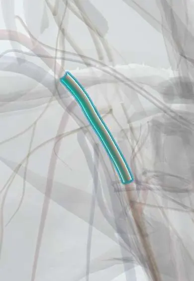
The axillary artery runs through the armpit; it feeds the shoulder and chest wall and continues as the brachial artery.
Opens in the Durgical app, no sign-up needed.
What it is
The main artery in the armpit, surrounded by the cords of the plexus.
What it does
Feeds the shoulder and continues as the brachial artery.
Key facts
- Origin
- Continuation of the subclavian artery at the 1st rib
- Branches
- 1st part: superior thoracic; 2nd: thoracoacromial, lateral thoracic; 3rd: subscapular, anterior and posterior circumflex humeral
- Supplies
- Shoulder, chest wall, axilla; becomes the brachial artery at the lower border of teres major
Relations
- Course
- From the outer border of the first rib it runs down and laterally through the axilla inside the axillary sheath with the brachial plexus, passing behind pectoralis minor, to the lower border of teres major
- In front
- Pectoralis major throughout; the clavipectoral fascia over the first part and pectoralis minor over the second; the lateral root of the median nerve crosses the third part
- Behind
- Posterior cord, then the radial nerve and axillary nerve; subscapularis, latissimus dorsi and teres major behind the third part
- Medial
- Axillary vein along its whole length; the medial cord, then the ulnar nerve and the medial cutaneous nerves between artery and vein
- Lateral
- Lateral cord, then the musculocutaneous nerve; coracobrachialis beside the third part
How to find it
Only its third part, below pectoralis minor, can be felt: with the arm abducted, the pulse is pressed against the humerus in the lower axilla just behind the bulge of coracobrachialis. On ultrasound for an axillary block, the median, ulnar and radial nerves surround the artery, the radial nerve usually deep to it.
On imaging
Duplex ultrasound and CT angiography are the usual tests. After an anterior shoulder dislocation or a proximal humeral fracture in an older patient, CT angiography looks for an intimal tear, occlusion or false aneurysm; the radiograph shows the bony injury that put it at risk. On ultrasound it is surrounded by the cords and branches of the brachial plexus, the landmark for an axillary block.
Clinical notes
- Anterior shoulder dislocation and proximal humerus fractures can stretch or tear its third part; older people with stiff, calcified arteries are at higher risk, including during reduction
- Because the cords hug it, arterial injury in the axilla often comes with a brachial plexus injury: a cold hand plus a nerve deficit after shoulder trauma suggests both
- Axillary brachial plexus block: the nerves surround the artery inside the sheath, so its pulse or its ultrasound image is the landmark
- The right axillary artery is a common cannulation site for cardiopulmonary bypass in aortic surgery
Anatomical variants
- Branching varies a lot: arterial variants are reported in roughly a quarter of upper limbs, and branches often share trunks, such as the subscapular with the circumflex humeral arteries
- High division: the radial or ulnar artery may arise from the axillary artery, giving two parallel arteries down the arm
Development
The arterial stem of the limb grows from the seventh cervical intersegmental artery: its first stretch becomes the subclavian, and its continuation into the bud forms the axillary and brachial arteries, which explains why these three are one continuous vessel.
Memory aid
English mnemonic for the branches in order, "Screw The Lawyer Save A Patient": Superior thoracic, Thoracoacromial, Lateral thoracic, Subscapular, Anterior and Posterior circumflex humeral (1, 2 and 3 per part).
Related conditions
Test yourself
Think of the answer, then tap the question to see it.
Which muscle divides the axillary artery into three parts, and how many branches does each give?
Pectoralis minor: above, behind and below it; the parts give one, two and three branches.
What are the branches of the axillary artery, by part?
First: superior thoracic; second: thoracoacromial and lateral thoracic; third: subscapular and anterior and posterior circumflex humeral.
Why does an axillary artery injury after shoulder trauma often come with a nerve deficit?
The brachial plexus cords hug the artery; a cold hand plus a nerve deficit suggests both are injured.
Why is an axillary artery block above the subscapular artery better tolerated than one below it?
Above it, flow is bypassed through the scapular anastomosis; below it, collateral flow is far poorer.
Educational information, not medical advice. For any symptom, see a health professional.
Its names
- Official Latin (TA2)
- arteria axillaris TA2 4616
- Official Spanish (DeCS)
- Arteria axilar
- English (TA2)
- Axillary artery
Related
Sources: Terminologia Anatomica (TA2); Anatomy, Shoulder and Upper Limb, Axillary Artery; Anatomy of the Human Body (20th ed.): Development of the Vascular System (Lea & Febiger (Gray's Anatomy, 1918), 1918) · adapted, Public domain; Anatomía humana (4.ª ed.), tomo 1, cap. 59: Arterias del miembro superior.
Spotted a mistake?We are always improving for the community. Tell us what is wrong and our team will review it.Report a mistake
Representative model, for educational purposes only.