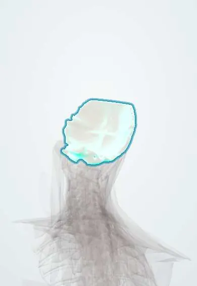
Base of the skull !
The occipital bone forms the back of the skull base, around the opening where the spinal cord meets the brain; it rests on the atlas.
Official name: Occipital bone
Opens in the Durgical app, no sign-up needed.
What it is
The back of the base of the skull, around the large hole (foramen magnum) where the spinal cord joins the brain.
What it does
Its two rounded knobs (condyles) sit on the atlas and form the 'yes' nodding joint.
Key facts
- Joints with
- Atlas via occipital condyles (atlanto-occipital joints); parietal and temporal bones (sutures); sphenoid (basilar part fuses with it)
- Landmarks
- Foramen magnum; occipital condyles; hypoglossal canals; clivus (basilar part); external occipital protuberance (inion); superior and inferior nuchal lines; pharyngeal tubercle (pharyngeal raphe); internal occipital protuberance (confluence of sinuses); internal occipital crest (falx cerebelli); condylar canals
Relations
- Above
- Posterior cranial fossa: the cerebellum on the inner squama, the brainstem on the clivus, and the transverse and sigmoid sinuses meeting near the internal occipital protuberance
- In front
- Basilar part (clivus) fused to the sphenoid; the pons, medulla and basilar artery rest on it inside, the nasopharynx lies below it outside
- Below
- Occipital condyles on the atlas (C1); suboccipital muscles, semispinalis and splenius insert between and below the nuchal lines
- Lateral
- Temporal bone at the occipitomastoid suture and the jugular foramen; the occipital artery and greater occipital nerve climb over the squama
- Contents
- Foramen magnum: the junction of medulla and spinal cord, both vertebral arteries, the spinal arteries and the spinal roots of the accessory nerve; the tectorial membrane and alar ligaments attach to its rim
How to find it
The external occipital protuberance (inion) is the midline bump at the back of the head; the superior nuchal line runs out from it, with trapezius attached near the midline and sternocleidomastoid laterally. The greater occipital nerve crosses the line a little lateral to the inion.
Examination
- Pressure or tapping over the greater occipital nerve where it crosses the superior nuchal line reproduces shooting pain up the back of the scalp in occipital neuralgia; relief after a local anaesthetic block there supports the diagnosis
On imaging
Sagittal MRI shows tonsil position in Chiari and the whole craniocervical junction; CT shows condyle fractures and basilar invagination, with the dens rising above reference lines such as Chamberlain line (hard palate to opisthion).
Clinical notes
- Occipital neuralgia: shooting pain over the back of the scalp along the greater occipital nerve; a block near the superior nuchal line is both test and treatment
- Decompression for symptomatic Chiari removes part of the squama around the foramen magnum, often with the posterior arch of C1
- Basilar invagination (rheumatoid arthritis, osteogenesis imperfecta, Paget disease) pushes the dens up into the foramen magnum against the brainstem
Anatomical variants
- Occipitalization of the atlas: partial or complete fusion with C1
- Interparietal (Inca) bone or small sutural (Wormian) bones along the lambdoid suture, which can mimic fractures in children
- Condylar canals and occipital emissary veins differ in size and may be absent on one or both sides
Development
Four parts around the foramen magnum (squama, two condylar parts and the basilar part) fuse in early childhood; the upper squama forms in membrane and the rest in cartilage. The basilar part fuses with the sphenoid in adolescence.
Related conditions
Test yourself
Think of the answer, then tap the question to see it.
What is a Chiari I malformation at the base of the skull (occipital bone)?
Descent of the cerebellar tonsils through the foramen magnum (commonly 5 mm or more below the basion–opisthion line), often with a syrinx and headache on coughing.
What passes through the hypoglossal canal and the jugular foramen at the base of the skull?
Hypoglossal canal: nerve XII. Jugular foramen (between occipital and temporal bones): nerves IX–XI and the internal jugular vein.
What passes through the foramen magnum of the occipital bone?
The junction of medulla and spinal cord, both vertebral arteries, the spinal arteries and the spinal roots of the accessory nerve.
Which nerve causes occipital neuralgia, and where is it blocked on the occipital bone?
The greater occipital nerve; a block near the superior nuchal line, a little lateral to the inion, is both test and treatment.
Educational information, not medical advice. For any symptom, see a health professional.
Its names
- Official Latin (TA2)
- os occipitale TA2 552
- Official Spanish (DeCS)
- Hueso occipital DeCS D009777
- English (TA2)
- Occipital bone
Related
- Alar ligaments
- Skull–atlas joints
- Longus capitis
- Nuchal ligament
- Rectus capitis anterior and lateralis
- Semispinalis
- Sternocleidomastoid
- Suboccipital muscles
Sources: Terminologia Anatomica (TA2); DeCS; Anatomy, Head and Neck, Occipital Bone, Artery, Vein, and Nerve; Anatomy, Head and Neck: Foramen Magnum.
Spotted a mistake?We are always improving for the community. Tell us what is wrong and our team will review it.Report a mistake
Representative model, for educational purposes only.