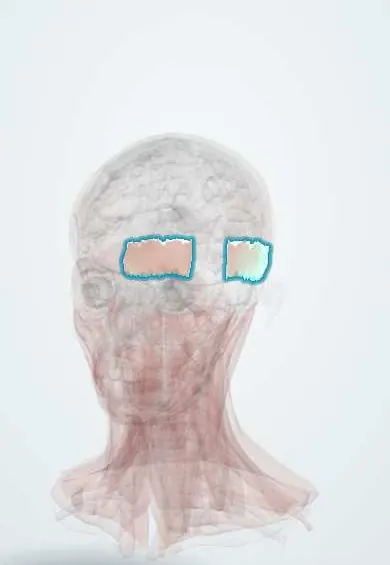
Muscular system · Head and neck
Occipitalis !
The occipitalis is a thin muscle at the back of the scalp; it pulls the scalp backwards, working with the forehead muscle.
Official name: Occipitalis muscle
Opens in the Durgical app, no sign-up needed.
What it is
A thin muscle at the back of the scalp.
What it does
Pulls the scalp back.
Key facts
- Origin
- Superior nuchal line, mastoid (some texts: highest nuchal line)
- Insertion
- Epicranial aponeurosis
- Action
- Pulls the scalp back
- Nerve
- Facial nerve (CN VII): posterior auricular nerve
- Blood supply
- Superficial temporal and occipital arteries
Relations
- Superficial
- Dense connective tissue and skin of the back of the scalp, with the occipital artery and the greater and lesser occipital nerves
- Deep
- Loose areolar tissue and pericranium on the occipital bone
- Above
- Epicranial aponeurosis (galea)
- Below
- Superior nuchal line, above the attachments of the trapezius and sternocleidomastoid
- Lateral
- Mastoid part of the temporal bone and the posterior auricular muscle behind the ear
How to find it
A thin quadrilateral sheet on each side of the back of the head, above the superior nuchal line; with fingers there, it is felt tensing as the eyebrows are raised hard and the scalp slides back.
On imaging
Rarely imaged on its own: it is a thin layer at the back of the scalp, over the occipital bone. Ultrasound and MRI show it among the scalp layers when a lump or haematoma is assessed. Nearby, ultrasound finds the greater occipital nerve and the occipital artery, the landmarks for an occipital nerve block in headache. In head trauma, CT shows soft-tissue swelling over the occiput.
Clinical notes
- Occipital neuralgia (shooting pain over the back of the head) involves the greater occipital nerve, which surfaces close to its lower medial border; nerve blocks target this point
- A cut through it and the galea at the back of the scalp gapes front to back, like a forehead wound
- The posterior auricular branch is spared in lesions inside the parotid, since it arises before the nerve enters the gland
Development
Second pharyngeal arch muscle, like the rest of the occipitofrontalis.
Related conditions
Test yourself
Think of the answer, then tap the question to see it.
Which branch of the facial nerve supplies the occipitalis?
The posterior auricular branch, which leaves the nerve just below the stylomastoid foramen, before the parotid.
Why is the occipitalis spared in lesions inside the parotid gland?
Its posterior auricular branch arises before the facial nerve enters the gland.
Which nerve involved in occipital neuralgia surfaces near the occipitalis?
The greater occipital nerve, close to its lower medial border; nerve blocks target this point.
Educational information, not medical advice. For any symptom, see a health professional.
Its names
- Official Latin (TA2)
- musculus occipitalis TA2 2057
- Official Spanish (DeCS)
- Vientre occipital del occipitofrontal
- English (TA2)
- Occipitalis muscle
- Also called
- occipital belly of epicranius muscle · occipital belly of occipitofrontalis muscle
Related
Sources: Terminologia Anatomica (TA2); Anatomy, Head and Neck; Frontalis Muscle; Anatomy, Head and Neck, Scalp.
Spotted a mistake?We are always improving for the community. Tell us what is wrong and our team will review it.Report a mistake
Representative model, for educational purposes only.