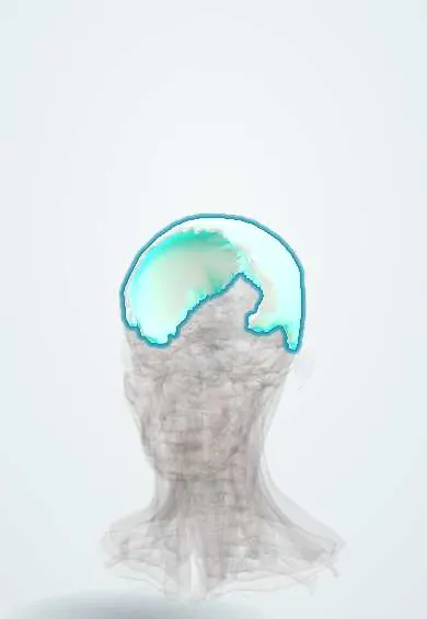
Skeletal system · Head and neck
Parietal bones !
The parietal bones are the two large curved bones forming the top and sides of the skull; they protect the upper part of the brain.
Official name: Parietal bone
Opens in the Durgical app, no sign-up needed.
What it is
The two large curved bones that form the top and sides of the skull.
What it does
Protect the top of the brain.
Key facts
- Joints with
- Opposite parietal (sagittal suture), frontal (coronal), occipital (lambdoid), temporal, sphenoid
- Landmarks
- Parietal eminence, temporal lines; groove for the middle meningeal artery inside
Relations
- Deep
- Dura mater with branches of the middle meningeal artery in grooves; the parietal lobe and parts of the frontal and occipital lobes; the superior sagittal sinus along the sagittal suture
- Superficial
- Scalp with the epicranial aponeurosis above; temporalis and its fascia from the temporal lines down
- Below
- Squamous part of the temporal bone at the squamous suture and, at the anteroinferior angle, the greater wing of the sphenoid bone at the pterion
- In front
- Frontal bone at the coronal suture
- Behind
- Occipital bone at the lambdoid suture
How to find it
The parietal eminence is the most prominent point on each side of the skull vault. In infants the anterior fontanelle is felt as a soft diamond at bregma and the smaller posterior fontanelle as a triangle at lambda.
On imaging
Non-contrast head CT: a biconvex hyperdense collection limited by sutures is extradural, a crescent that crosses sutures is subdural. Bone windows show linear or depressed fractures, which are not to be confused with sutures or vascular grooves.
Clinical notes
- In newborns, a cephalohaematoma is a bleed under the periosteum, most often over a parietal bone; it stops at the suture lines, unlike caput succedaneum
- Fusion of the sagittal suture, the most common single-suture craniosynostosis, gives a long, narrow head (scaphocephaly)
- The outer table of the parietal bone is a common donor site for split calvarial bone grafts in facial reconstruction
Anatomical variants
- Small sutural (Wormian) bones along the lambdoid suture are common; large numbers of them can point to a bone disorder such as osteogenesis imperfecta
- Enlarged parietal foramina, a rare inherited defect felt as symmetrical soft spots
Development
Ossifies in membrane from the region of the parietal eminence. The posterior fontanelle usually closes within the first 2 to 3 months, the anterior by about 18 to 24 months.
Related conditions
- Multiple myeloma
- Child maltreatment (physical abuse, abusive head trauma, neglect)
- Neonatal birth injuries (caput, cephalohaematoma, clavicle fracture)
- Epidural haematoma
Test yourself
Think of the answer, then tap the question to see it.
Which artery grooves the inner surface of the parietal bone, and what does tearing it cause?
Branches of the middle meningeal artery; a fracture at the pterion or anteroinferior angle can tear them and cause an extradural (epidural) haematoma.
What are the classic clinical course and CT appearance of an extradural haematoma?
Brief loss of consciousness, a lucid interval, then rapid decline; on CT a biconvex clot that does not cross sutures.
Which fontanelles lie at bregma and at lambda on the parietal bones?
The anterior fontanelle at bregma (coronal meets sagittal suture) and the posterior fontanelle at lambda (sagittal meets lambdoid suture).
How does a cephalohaematoma over a parietal bone differ from caput succedaneum in a newborn?
A cephalohaematoma is a bleed under the periosteum, so it stops at the suture lines; caput succedaneum does not.
Educational information, not medical advice. For any symptom, see a health professional.
Its names
- Official Latin (TA2)
- os parietale TA2 504
- Official Spanish (DeCS)
- Hueso Parietal DeCS D010294
- English (TA2)
- Parietal bone
Sources: Terminologia Anatomica (TA2); DeCS; Anatomy, Head and Neck, Skull; Anatomy, Head and Neck, Coronal Suture.
Spotted a mistake?We are always improving for the community. Tell us what is wrong and our team will review it.Report a mistake
Representative model, for educational purposes only.