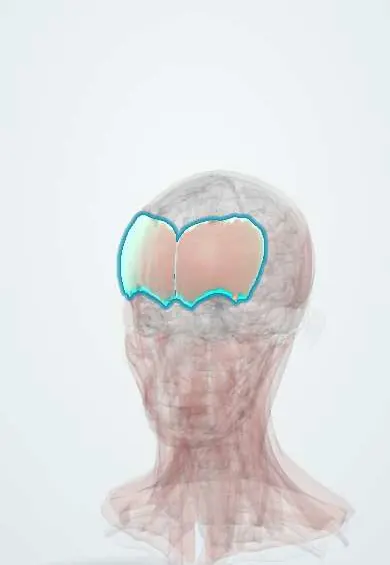
Muscular system · Head and neck
Frontalis !
The frontalis is the thin muscle of the forehead; it raises the eyebrows and wrinkles the forehead, and goes slack on one side in Bell's palsy.
Official name: Occipitofrontalis muscle
Opens in the Durgical app, no sign-up needed.
What it is
The thin muscle of the forehead.
What it does
Raises the eyebrows and wrinkles the forehead; in Bell's palsy one side of the forehead goes smooth.
Key facts
- Origin
- Epicranial aponeurosis
- Insertion
- Skin of the eyebrows and forehead
- Action
- Raises the eyebrows, wrinkles the forehead
- Nerve
- Facial nerve (CN VII): temporal branches
- Blood supply
- Superficial temporal and occipital arteries
Relations
- Superficial
- Skin and dense subcutaneous tissue of the forehead, with the supraorbital and supratrochlear nerves and vessels after they pierce the muscle
- Deep
- Loose areolar tissue and the pericranium over the frontal bone; the supraorbital notch and supratrochlear vessels at the brow
- Above
- Epicranial aponeurosis (galea)
- Below
- Interlaces with the procerus, corrugator supercilii and orbicularis oculi at the brow
How to find it
Asked to raise the eyebrows and look up, horizontal forehead creases appear over the two muscle bellies, which are separated by a thin gap in the midline.
Examination
- Raising the eyebrows: no forehead creases on one side means a peripheral (lower motor neuron) lesion of the facial nerve, as in Bell palsy; creases preserved with a weak lower face suggest a central lesion such as a stroke
- The House–Brackmann scale grades facial nerve function partly by forehead movement
On imaging
Rarely imaged: facial nerve function is judged clinically. Ultrasound shows it as a thin layer under the forehead skin, used to map it before botulinum toxin and to place a forehead lump; lipomas here often lie under the muscle. In facial palsy, MRI studies the nerve, not the muscle. In forehead trauma, CT shows scalp swelling over the frontal bone and any fracture of the frontal sinus.
Clinical notes
- Temporal branch injury causes a drooping brow that does not lift; its course is often drawn along Pitanguy's line, from about 0.5 cm below the tragus to about 1.5 cm above the lateral end of the eyebrow
- Botulinum toxin injection for forehead lines is a common procedure; too much weakening can drop the brow and make the upper lids feel heavy
- The frontalis sling operation connects a drooping eyelid to this muscle when the levator palpebrae superioris works poorly, as in some congenital ptosis
Development
Second pharyngeal arch muscle; with the occipitalis it forms the occipitofrontalis.
Memory aid
Facial nerve branches from top to bottom: «To Zanzibar By Motor Car» (temporal, zygomatic, buccal, marginal mandibular, cervical); the frontalis takes the first, T.
Related conditions
Test yourself
Think of the answer, then tap the question to see it.
Why is the forehead spared in a central (upper motor neuron) facial palsy but not in Bell palsy?
The upper face gets cortical input from both hemispheres, so the frontalis still wrinkles the forehead in a central lesion.
Which branch of the facial nerve supplies the frontalis, and what does its injury cause?
The temporal (frontal) branch, crossing the zygomatic arch under the temporoparietal fascia; injury gives a drooping brow that does not lift.
What is the frontalis sling operation?
Connecting a drooping eyelid to the frontalis when the levator palpebrae superioris works poorly, as in some congenital ptosis.
Educational information, not medical advice. For any symptom, see a health professional.
Its names
- Official Latin (TA2)
- musculus occipitofrontalis TA2 2055
- Official Spanish (DeCS)
- Vientre frontal del músculo occipitofrontal DeCS D005152
- English (TA2)
- Occipitofrontalis muscle
Related
Sources: Terminologia Anatomica (TA2); DeCS; Anatomy, Head and Neck; Frontalis Muscle; Anatomy, Head and Neck: Facial Muscles.
Spotted a mistake?We are always improving for the community. Tell us what is wrong and our team will review it.Report a mistake
Representative model, for educational purposes only.