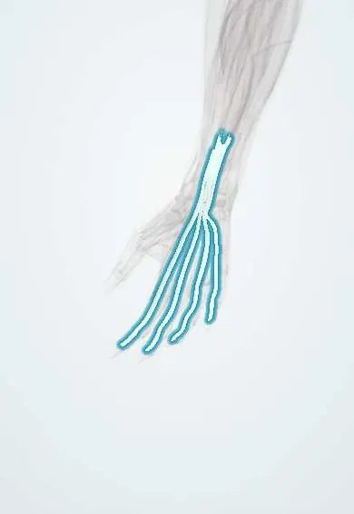
Flexor tendon injury !
A flexor tendon injury is a cut of the tendons that bend the fingers or thumb, usually from a wound on the palm side of the hand, wrist or forearm.
Updated 2026-10-29
Structures involved
Tap one to see what it is and open it in 3D.
- FDP tendons
- FDS tendons
- Flexor tendon sheaths
- Finger pulleys
- FPL
- A1 pulleys
- Digital nerves
- Digital arteries
- FDP
- FDS
Why it happens
- Tissue repair: regeneration or scar; labile, stable and permanent cells
- Connective tissue proper: cells and fibres
What it is
Partial or complete division of the tendons that bend the fingers and thumb (flexor digitorum profundus, flexor digitorum superficialis and flexor pollicis longus), usually by a cut on the palm side of the hand, wrist or forearm. Treatment and outcome depend on the anatomical zone of the injury.
Who gets it
Mostly young working-age adults, more often men, after injuries with knives, glass, tools and machinery at home or at work. Closed avulsion of the profundus tendon (jersey finger) occurs in contact sports.
Causes
- Cuts from knives, glass, metal sheets or tools
- Crush and machinery injuries
- Closed avulsion of the profundus tendon from the distal phalanx when a flexed finger is forced straight (jersey finger, usually the ring finger)
- Attritional rupture in rheumatoid arthritis or over bony prominences
Risk factors
- Manual work with sharp tools or machinery
- Kitchen and glass injuries
- Contact sports such as football and rugby (closed avulsion)
- Rheumatoid arthritis
Pathophysiology
In the fingers the two flexor tendons run inside a tight synovial sheath held to the bone by pulleys, of which A2 and A4 prevent bowstringing. Cut tendons retract and do not heal unless their ends are brought together; their nutrition comes from vincula and synovial fluid. Healing scar easily binds tendon to sheath, so tight repairs with early protected movement are needed to keep gliding.
Classification
- Zone I: distal to the superficialis insertion, profundus tendon only
- Zone II (no man's land): from the distal palmar crease (A1 pulley) to the superficialis insertion, both tendons inside the tight sheath
- Zone III: palm, where the lumbricals arise
- Zone IV: within the carpal tunnel
- Zone V: wrist and forearm, proximal to the carpal tunnel
- Thumb zones T I to T V; jersey finger avulsions are graded by Leddy and Packer
Symptoms and signs
- Inability to bend one or more finger joints
- Loss of the normal resting flexion cascade: the injured finger lies straighter than its neighbours
- Abnormal tenodesis effect: the finger does not flex when the wrist is passively extended
- Numbness of the side of a finger if the digital nerve is also cut
- Wound on the palm side of the finger, palm or wrist
Diagnosis
- Clinical
- Profundus test: with the middle joint held straight, the patient cannot bend the end joint. Superficialis test: with the other fingers held straight, the patient cannot bend the middle joint. Sensation and capillary refill of each digit are checked.
- Tests
- Not usually needed; tetanus status is reviewed.
- Imaging
- X-rays look for fractures, bony avulsion and glass or metal fragments; ultrasound or MRI can locate a retracted tendon end or a partial tear.
Differential diagnosis
- Anterior interosseous or median nerve palsy
- Trigger finger with locking
- Pulley rupture
- Joint stiffness or fracture limiting flexion
- Pain inhibition without tendon injury
Goal of treatment
Restore continuity of the tendon with a repair strong enough for early protected movement, preserve the key pulleys and regain smooth gliding and full finger motion.
Treatment
- Acute
- Wound cleaning, closure or dressing, splint with wrist and fingers flexed, tetanus prophylaxis and antibiotics for contaminated wounds, and referral to hand surgery.
- Surgical
- Primary repair, usually within the first days, with multi-strand core sutures and an epitendinous suture, preserving the A2 and A4 pulleys and repairing cut nerves and arteries.
- Surgical
- Jersey finger: re-attachment of the profundus tendon to the distal phalanx, urgent when the tendon has retracted into the palm.
- Rehabilitation
- Hand therapy with a protective splint and early controlled active or passive motion protocols for several weeks, then strengthening.
- Surgical
- Late or failed cases: tenolysis for adhesions, or staged tendon reconstruction with grafts.
Complications
- Adhesions that limit tendon gliding
- Re-rupture of the repair
- Joint contracture and stiffness
- Bowstringing after pulley damage
- Infection and quadriga effect (limited flexion of neighbouring fingers)
Prognosis
Outcome depends on the zone, the cleanliness of the wound, the quality of repair and adherence to therapy. Zone II injuries have historically had the most adhesions; modern repair with early motion has improved results, but full motion is not always regained.
Prevention
- Safe handling of knives, glass and tools
- Machine guards and gloves at work
Test yourself
Think of the answer, then tap the question to see it.
Why is flexor tendon zone II called no man's land?
Both flexor tendons run together inside the tight fibro-osseous sheath, where repairs easily form adhesions and historically had poor results.
How is the flexor digitorum profundus tested in a finger?
The examiner holds the PIP joint straight; inability to flex the DIP joint indicates a cut profundus tendon.
How is the flexor digitorum superficialis tested in a finger?
The other fingers are held in extension to block the profundus; inability to flex the PIP joint indicates a cut superficialis tendon.
Which pulleys are most important to preserve to prevent bowstringing?
The A2 and A4 pulleys.
What is a jersey finger?
Closed avulsion of the profundus tendon from the distal phalanx when a flexed finger is forcibly extended, most often the ring finger.
Related conditions
Educational information, not medical advice. For any symptom, see a health professional.
Sources: Flexor Tendon Lacerations (StatPearls (NCBI Bookshelf), 2023).
Spotted a mistake?We are always improving for the community. Tell us what is wrong and our team will review it.Report a mistake
Representative model, for educational purposes only.