Nervous system · Head and neck
Globus pallidus !
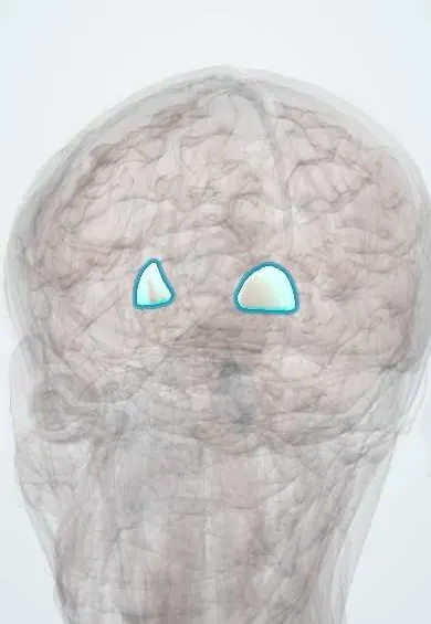
The globus pallidus is the pale inner part of a lens-shaped mass deep in the brain; it is the main output of the basal ganglia that shape movement.
Opens in the Durgical app, no sign-up needed.
What it is
The pale inner part of the lens-shaped nucleus.
What it does
The main output of the basal ganglia; a target of deep brain stimulation.
Key facts
- Where
- Medial part of the lentiform nucleus
- Blood supply
- Anterior choroidal artery (mainly the internal segment); lenticulostriate arteries from the MCA (mainly the external segment)
- Function
- Main output of the basal ganglia (DBS target)
Relations
- Lateral
- Putamen, separated by the lateral medullary lamina; together they make the lentiform nucleus
- Medial
- Genu and posterior limb of the internal capsule, and beyond them the thalamus
- Below
- Across the internal capsule below and medially, the subthalamic nucleus and the substantia nigra; the pallidal output fibres (ansa and fasciculus lenticularis) loop around and through the capsule to reach the thalamus
How to find it
It is the inner, paler part of the lentiform wedge, medial to the putamen and lateral to the genu and posterior limb of the internal capsule; a thin white lamina divides its external and internal segments. In older adults it often looks dark on T2-weighted MRI because of normal iron deposits.
On imaging
Pale on fresh brain because myelinated fibres cross it. On CT, calcification of both pallidi is a common harmless finding in older adults. On MRI, symmetric T2-bright pallidi suggest carbon monoxide poisoning; T1-bright pallidi are seen in chronic liver disease (manganese deposits); a dark pallidus with a bright centre ('eye of the tiger') suggests pantothenate kinase-associated neurodegeneration.
Clinical notes
- Its neighbour, the subthalamic nucleus, drives the internal pallidus; a small stroke there releases the thalamus and causes hemiballismus: wild flinging movements of the opposite limbs
- Pallidotomy or internal pallidus stimulation eases the involuntary movements (dyskinesias) that can follow long-term levodopa treatment
- Kernicterus: in newborns with very high bilirubin, the pigment stains and injures the globus pallidus, leading to dystonia and choreoathetosis
Memory aid
Basal ganglia in two words: the direct pathway says 'go', the indirect says 'stop'; dopamine presses 'go' and lifts 'stop'.
Related conditions
- Methylmalonic acidaemia
- Propionic acidaemia
- Guanidinoacetate methyltransferase (GAMT) deficiency
- Cyanide poisoning
- Carbon monoxide poisoning
- Parkinson disease
- Dystonia (generalised and focal)
- Antipsychotic-induced movement disorders (dystonia, akathisia, parkinsonism, tardive dyskinesia)
- Catatonia
Test yourself
Think of the answer, then tap the question to see it.
What is the role of the internal globus pallidus in the basal ganglia?
With the substantia nigra pars reticulata, it is the inhibitory output to the thalamus, the main output of the basal ganglia.
Which poisoning damages both globi pallidi symmetrically?
Carbon monoxide poisoning (as does severe hypoxia); MRI shows symmetric T2-bright pallidi.
Which neighbouring nucleus, when damaged, releases the thalamus and causes hemiballismus?
The subthalamic nucleus, which drives the internal pallidus; a small stroke there gives wild flinging movements of the opposite limbs.
What is kernicterus, and which part of the brain does it injure?
Very high bilirubin in newborns stains and injures the globus pallidus, leading to dystonia and choreoathetosis.
Educational information, not medical advice. For any symptom, see a health professional.
Its names
- Official Latin (TA2)
- globus pallidus TA2 5569
- Official Spanish (DeCS)
- Globo Pálido DeCS D005917
- English (TA2)
- Globus pallidus
- Also called
- dorsal pallidum · paleostriatum
Related
Sources: Terminologia Anatomica (TA2); DeCS; Neuroanatomy, Globus Pallidus; 13.2 The Central Nervous System (OpenStax (Rice University), 2022) · adapted, CC BY 4.0.
Spotted a mistake?We are always improving for the community. Tell us what is wrong and our team will review it.Report a mistake
Representative model, for educational purposes only.