Nervous system · Head and neck
Putamen !
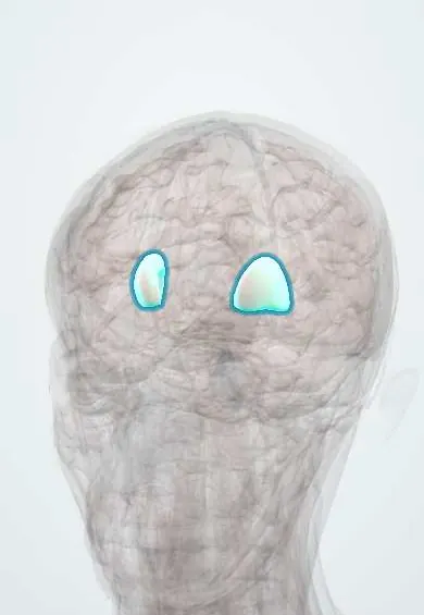
The putamen is the outer part of a lens-shaped mass deep in each half of the brain; it helps control movement and often bleeds with high blood pressure.
Opens in the Durgical app, no sign-up needed.
What it is
The outer part of the lens-shaped nucleus deep in each hemisphere.
What it does
Basal ganglia motor loop; a common site of high-blood-pressure bleeds.
Key facts
- Where
- Lateral part of the lentiform nucleus
- Blood supply
- Lenticulostriate arteries (MCA)
- Function
- Motor loop of the basal ganglia; common hypertensive bleed site
Relations
- Lateral
- External capsule, claustrum, extreme capsule and then the cortex of the insula
- Medial
- Globus pallidus, separated by a thin sheet of white matter (lateral medullary lamina); beyond it the internal capsule
- In front
- Joined to the head of the caudate nucleus by grey bridges across the anterior limb of the internal capsule and, below, at the nucleus accumbens
- Above
- Corona radiata of the white matter, through which the lenticulostriate arteries climb from the middle cerebral artery
How to find it
On axial and coronal images the lentiform nucleus is a wedge with its broad base facing out; the putamen is its outer part, lateral to the globus pallidus, with the internal capsule medial and the external capsule, claustrum and insula lateral. Above and in front it joins the caudate head through grey bridges across the capsule.
On imaging
A hypertensive bleed shows on CT as a bright (hyperdense) mass lateral to the internal capsule. On dopamine transporter SPECT the striatum looks like a comma; in Parkinson disease uptake fades first in the back of the putamen, leaving a 'full stop'. Bilateral putaminal necrosis or bleeding is a classic finding in methanol poisoning.
Clinical notes
- Wilson disease: copper builds up in the lentiform nucleus (hence 'hepatolenticular degeneration'), causing tremor, dystonia and rigidity
- Large putaminal bleeds can extend into the ventricles or push the brain across the midline; the depth of the putamen is why surgery for most of them is not routine
- Lacunar infarcts from blocked lenticulostriate arteries here and in the internal capsule cause pure motor strokes
Development
Caudate and putamen begin as one grey mass in the floor of the lateral ventricle (the ganglionic eminence); the fibres of the internal capsule grow through it and split it, leaving the grey strands that give the striatum its striped look.
Memory aid
Putamen is Latin for 'shell' or 'husk': it is the outer shell of the lentiform nucleus, wrapped around the globus pallidus.
Related conditions
- Pyruvate dehydrogenase deficiency
- Huntington disease
- Lesch-Nyhan syndrome
- Cyanide poisoning
- Leigh syndrome
- Methanol poisoning
- Wilson disease
- Intracerebral haemorrhage
- Parkinson disease
- Dystonia (generalised and focal)
- Multiple system atrophy (MSA)
- Perinatal asphyxia and hypoxic-ischaemic encephalopathy
- Stimulant use disorder (cocaine, amphetamines)
- Antipsychotic-induced movement disorders (dystonia, akathisia, parkinsonism, tardive dyskinesia)
- Tourette syndrome and tic disorders
Test yourself
Think of the answer, then tap the question to see it.
What is the most common site of hypertensive brain haemorrhage, and why?
The putamen: a lenticulostriate artery ruptures and the clot spreads into the internal capsule.
What are the signs of a putaminal haemorrhage?
Opposite-side weakness and numbness, with the eyes turned toward the bleed.
How does dopamine loss in Parkinson disease affect the putamen's pathways?
Dopamine normally boosts the direct («go») pathway and damps the indirect («stop») one; its loss leaves the brake on, so movement becomes slow and scant.
Which metal builds up in the lentiform nucleus in Wilson disease?
Copper, hence «hepatolenticular degeneration», causing tremor, dystonia and rigidity.
Educational information, not medical advice. For any symptom, see a health professional.
Its names
- Official Latin (TA2)
- putamen TA2 5566
- Official Spanish (DeCS)
- Putamen DeCS D011699
- English (TA2)
- Putamen
Related
Sources: Terminologia Anatomica (TA2); DeCS; Neuroanatomy, Putamen; 13.2 The Central Nervous System (OpenStax (Rice University), 2022) · adapted, CC BY 4.0.
Spotted a mistake?We are always improving for the community. Tell us what is wrong and our team will review it.Report a mistake
Representative model, for educational purposes only.