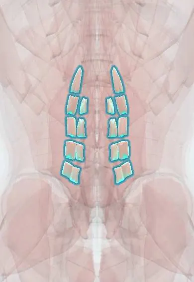
Intertransversarii !
The intertransversarii are small muscles between neighbouring transverse processes; they help bend the spine sideways.
Official name: Intertransversarii muscles
Opens in the Durgical app, no sign-up needed.
What it is
Small muscles between neighbouring transverse processes.
What it does
Help bend the spine sideways and sense its position.
Key facts
- Origin
- Transverse process below (cervical: anterior and posterior slips; lumbar: medial and lateral slips)
- Insertion
- Transverse process of the vertebra above
- Action
- Assist lateral flexion; stabilise; proprioception
- Nerve
- Dorsal and ventral rami (posterior cervical and medial lumbar: dorsal rami; anterior cervical and lateral lumbar: ventral rami)
- Blood supply
- Segmental arteries: vertebral and deep cervical, posterior intercostal, lumbar
Relations
- Medial
- Neck: the vertebral artery climbing through the transverse foramina; lumbar: the facet joints and multifidus
- Lateral
- Lumbar: quadratus lumborum and psoas major, attached to the same transverse processes
- Contents
- Between the cervical slips: the ventral ramus of each spinal nerve, which has just crossed behind the vertebral artery; in the lumbar region the intertransverse membrane with the exiting nerve root in front of it
How to find it
They cannot be felt. In a far-lateral (extraforaminal) lumbar approach they are the thin muscle between two transverse processes, divided with the intertransverse membrane to reach the exiting nerve root.
On imaging
Not seen separately on routine imaging; axial and coronal MRI show the thin soft tissue between neighbouring transverse processes, just outside the foramen. What is assessed there is the exiting nerve root: in a far-lateral (extraforaminal) disc herniation, MRI shows disc material lateral to the foramen pressing on the root in this space. CT shows transverse process fractures nearby.
Common problems
- Shrinks and fills with fat in chronic low back pain, sometimes only at the painful level.
- Is stripped or split from the bone in open posterior spine surgery; muscle-sparing approaches try to protect it.
How it is diagnosed
MRI or ultrasound shows its size and fat content.
Treatment
- Targeted exercise (motor control training) to retrain the deep stabilisers.
Clinical notes
- Far-lateral lumbar disc herniation: the muscle and the intertransverse membrane are opened between two transverse processes to reach the compressed exiting root
- In lateral exposures of the cervical spine the vertebral artery and the ventral rami lie right at their slips
Development
Short segmental muscles that keep the one-level pattern of the myotomes. Their split nerve supply reflects a double origin: the medial lumbar slips belong to the back (dorsal rami), while the lateral lumbar and anterior cervical slips develop with the body wall (ventral rami).
Test yourself
Think of the answer, then tap the question to see it.
What emerges between the anterior and posterior cervical slips of the intertransversarii?
The ventral ramus of each cervical spinal nerve.
How are the lumbar intertransversarii supplied?
Medial slips by dorsal rami, lateral slips by ventral rami.
Why are the intertransversarii opened in a far-lateral lumbar disc herniation?
Opening them and the intertransverse membrane between two transverse processes reaches the compressed exiting nerve root.
Educational information, not medical advice. For any symptom, see a health professional.
Its names
- Official Latin (TA2)
- musculi intertransversarii TA2 2294
- Official Spanish (DeCS)
- Músculos intertransversos DeCS D064170
- English (TA2)
- Intertransversarii muscles
Related
Sources: Terminologia Anatomica (TA2); DeCS; Anatomy, Back; Anatomy of the Human Body (20th ed.): Development of the Muscles (Lea & Febiger (Gray's Anatomy, 1918), 1918) · adapted, Public domain; Anatomía humana (4.ª ed.), tomo 1, cap. 19: Músculos del cuello.
Spotted a mistake?We are always improving for the community. Tell us what is wrong and our team will review it.Report a mistake
Representative model, for educational purposes only.