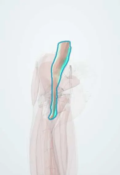
Psoas major !
The psoas major is a long muscle running from the lower spine to the femur; it is the main muscle that lifts the thigh.
Opens in the Durgical app, no sign-up needed.
What it is
A long, thick muscle that starts on the sides of the lower spine, runs through the pelvis and joins the iliacus to attach to the lesser trochanter, a bump on the inner top of the femur.
What it does
The main hip flexor: lifts the thigh when you walk, run or climb stairs, and helps steady the lower spine. With the iliacus it is called the iliopsoas.
Key facts
- Origin
- Sides of T12–L5 vertebral bodies and discs; transverse processes of L1–L5
- Insertion
- Lesser trochanter of femur (with iliacus, as iliopsoas)
- Action
- Flexes hip (main hip flexor with iliacus); flexes trunk; lateral flexion of lumbar spine
- Nerve
- Anterior rami of lumbar nerves (L1–L3) (some texts: L1–L4 or L2–L4)
- Blood supply
- Lumbar arteries; iliolumbar artery (lumbar branch)
Relations
- Medial
- Lumbar vertebral bodies and the sympathetic trunk; the abdominal aorta on the left, the inferior vena cava on the right
- In front
- Kidney and ureter, gonadal vessels and genitofemoral nerve; caecum and appendix on the right, descending and sigmoid colon on the left
- Lateral
- Quadratus lumborum above, iliacus below, with the femoral nerve in the groove between the psoas and the iliacus
- Behind
- Transverse processes of the lumbar vertebrae
- Above
- Medial arcuate ligament of the diaphragm, which arches over its upper end
- Course
- Runs down along the pelvic brim, passes under the inguinal ligament with the iliacus and the femoral nerve, crosses the front of the hip joint capsule and turns back to the lesser trochanter
- Contents
- Roots and first branches of the lumbar plexus
How to find it
Deep: on CT or MRI it is the paired muscle beside the lumbar vertebral bodies, widening as it descends. In the groin, as the iliopsoas, it forms the lateral part of the floor of the femoral triangle, under the femoral nerve.
Examination
- Psoas sign: lying on the left side, the right hip is extended passively (or flexed against resistance); pain suggests retrocaecal appendicitis or another inflammatory process against the psoas
- Resisted hip flexion while seated tests mainly the L2 myotome; weakness can come from the muscle itself, the upper lumbar roots or the lumbar plexus
On imaging
Contrast CT is the usual test for a psoas abscess: an enlarged muscle with a low-density collection and an enhancing rim, sometimes with the infected vertebra and disc next to it.
Common problems
- Strain or tendinitis from kicking, sprinting or repeated hip bending.
- Snapping hip (inside type): the tendon catches over the front of the pelvis with a clunk.
- Tight hip flexors from long sitting can add to back or hip pain.
- Uncommonly, tendon irritation after hip replacement if it rubs on the implant.
How it is diagnosed
Physical exam (pain when lifting the leg against resistance); ultrasound, which can also show the snapping during movement; MRI.
Treatment
- Rest from the painful activity, stretching and progressive strengthening with a physical therapist.
- Ultrasound-guided injection into the iliopsoas bursa or around the tendon.
- Rarely, surgery to lengthen or release the tendon when snapping or pain does not improve.
Clinical notes
- Irritation of the psoas makes the person keep the hip flexed, because extension stretches the inflamed muscle
- Lateral transpsoas approaches to the lumbar spine pass through it and can injure the lumbar plexus or the genitofemoral nerve, causing thigh numbness or hip flexor weakness
Anatomical variants
- The psoas minor, a thin muscle in front of the psoas major, is missing in many people: on both sides in about 2 in 5 in the cited review
- A double (bifid) iliopsoas tendon is not rare: on one side in about 1 in 4 in the cited review
Related conditions
- Pyomyositis (including psoas abscess)
- Intra-abdominal abscess
- Acute appendicitis
- Kidney stones (urolithiasis)
- Acute pyelonephritis
- Intertrochanteric hip fracture
Test yourself
Think of the answer, then tap the question to see it.
Where do the femoral and obturator nerves leave the psoas major?
The femoral nerve leaves its lateral border and the obturator nerve its medial border; the genitofemoral nerve pierces its front.
Where does the psoas major insert, and what is its main action?
On the lesser trochanter of the femur, with the iliacus as the iliopsoas; it is the main hip flexor.
What does a positive psoas sign suggest?
Retrocaecal appendicitis or an abscess against the psoas: pain on passive hip extension (or resisted flexion) with the patient lying on the left side.
How can a psoas abscess from the spine present in the groin?
It tracks down the psoas sheath (classically from tuberculous spondylitis) and can appear below the inguinal ligament.
What is the nerve supply of the psoas major?
Anterior rami of the lumbar nerves, L1–L3 (some texts: L1–L4 or L2–L4).
Educational information, not medical advice. For any symptom, see a health professional.
Its names
- Official Latin (TA2)
- psoas major TA2 2595
- Official Spanish (DeCS)
- Músculo psoas mayor DeCS D016658
- English (TA2)
- Psoas major
- Also called
- psoas major muscle · musculus psoas major
Related
Sources: Terminologia Anatomica (TA2); DeCS; Anatomy, Bony Pelvis and Lower Limb: Psoas Major; Anatomy, Bony Pelvis and Lower Limb: Iliopsoas Muscle.
Spotted a mistake?We are always improving for the community. Tell us what is wrong and our team will review it.Report a mistake
Representative model, for educational purposes only.