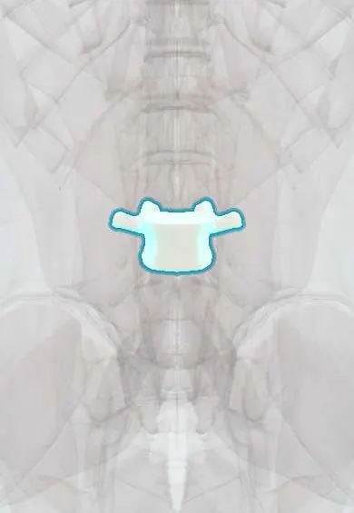
L3 vertebra !
L3 is the third lumbar (lower-back) vertebra; its large body carries the weight of the upper body and lets the lower back bend.
Official name: L3
Opens in the Durgical app, no sign-up needed.
What it is
The L3 lower-back vertebra, with a large body built to carry weight.
What it does
Carries the weight of the upper body and, with the disc and facet joints, lets the lower back bend forward and back.
Key facts
- Joints with
- L2 above and L4 below (intervertebral discs, facet joints)
- Landmarks
- Large kidney-shaped body; no costal facets or transverse foramina; mammillary processes; usually the longest transverse processes; umbilicus near L3–L4 disc
Relations
- In front
- Third part of the duodenum crossing between the superior mesenteric artery and the abdominal aorta; the inferior mesenteric artery leaves the aorta; the inferior vena cava to the right
- Lateral
- Psoas major with the lumbar plexus; the right crus of the diaphragm reaches down to this body; the lower poles of the kidneys lie near this level
- Behind
- Quadratus lumborum and the deep layers of the thoracolumbar fascia attach to the long transverse processes; erector spinae behind
- Contents
- Cauda equina in the dural sac; the L3–L4 gap is a usual lumbar puncture level in adults
How to find it
The L3 spine lies one level above the L4 spine found on the line joining the tops of the iliac crests. In slim people the umbilicus is a rough guide in front.
Examination
- Patellar (knee) reflex, mainly L4 with L3: reduced with an L3–L4 herniation
On imaging
On the AP lumbar radiograph L3 usually has the longest transverse processes; after trauma CT shows transverse process fractures and should prompt a look for injury to nearby organs. On axial CT the L3 slice is the standard level for measuring skeletal muscle area in sarcopenia. MRI is chosen for discs, nerve roots, marrow and infection.
Common problems
- Disc degeneration and herniation at L4-L5, the most common level for back and leg pain.
- Degenerative spondylolisthesis: L4 slips forward on L5, often with stenosis (more common in women over 50).
- Spinal stenosis: the canal narrows from disc bulging, facet arthritis and a thick ligamentum flavum.
- Compression fractures from osteoporosis or injury.
How it is diagnosed
X-rays (standing, and bending forward and back) show alignment and slipping. MRI shows discs, nerves and stenosis. CT shows bone detail.
Treatment
- Most back pain improves with staying active, physical therapy and pain medicine.
- Stenosis with leg pain when walking that does not improve: decompression (laminectomy).
- Slipping with instability: decompression plus fusion with screws, rods, a cage and bone graft.
Clinical notes
- Superior mesenteric artery syndrome: the third part of the duodenum is squeezed between the SMA and the aorta in front of L3, typically after rapid weight loss; vomiting after meals
- Fractures of the long L3 transverse processes are stable for the spine but signal high-energy trauma, with possible kidney or bowel injury
- Abdominal aortic aneurysms usually lie between the renal arteries and the bifurcation, in front of L2–L4, and are felt as a pulsatile mass above the umbilicus
Development
The lumbar lordosis is a secondary curve: it forms as the child begins to stand and walk, and its apex is usually around L3. It is generally deeper in women.
Related conditions
Test yourself
Think of the answer, then tap the question to see it.
Which plane crosses the L3 vertebra?
The subcostal plane, through the lowest points of the costal margins (tenth costal cartilages).
Which part of the bowel crosses in front of the L3 vertebra, and what syndrome can compress it?
The third part of the duodenum; in superior mesenteric artery syndrome it is squeezed between the SMA and the aorta, typically after rapid weight loss.
Which nerve root does an L3–L4 posterolateral disc herniation compress, and what are the signs?
The L4 nerve root: numb medial leg, weak knee extension and a reduced patellar reflex.
What does a fracture of the long transverse processes of the L3 vertebra signal?
It is stable for the spine but signals high-energy trauma, with possible kidney or bowel injury.
Educational information, not medical advice. For any symptom, see a health professional.
Its names
- Official Latin (TA2)
- vertebra lumbalis III TA2 1068
- Official Spanish (DeCS)
- Vértebra lumbar L3 DeCS D008159
- English (TA2)
- L3
Related
- L2–L3 disc
- L3–L4 disc
- Lumbar arteries
- Anterior longitudinal ligament
- Diaphragm
- Endplates
- Low-back facet joints
- Iliopsoas fascia
Sources: Terminologia Anatomica (TA2); DeCS; Anatomy, Back, Lumbar Spine; 7.3 The Vertebral Column (OpenStax (Rice University), 2022) · adapted, CC BY 4.0.
Spotted a mistake?We are always improving for the community. Tell us what is wrong and our team will review it.Report a mistake
Representative model, for educational purposes only.