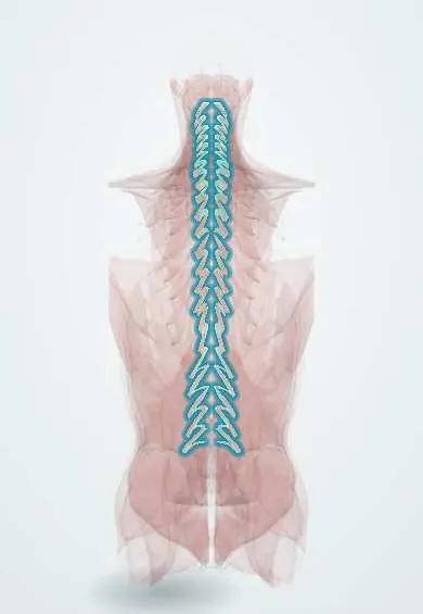
Multifidus !
The multifidus is a deep back muscle of many short bundles, thickest in the lower back; it holds each vertebra steady during movement.
Official name: Multifidus muscles
Opens in the Durgical app, no sign-up needed.
What it is
A deep back muscle made of many short bundles, each crossing two to four vertebrae, thickest in the lower back.
What it does
Holds each vertebra steady during movement (a key stabiliser) and helps straighten and twist the spine.
Key facts
- Origin
- Sacrum, posterior superior iliac spine, erector spinae aponeurosis, lumbar mammillary processes, thoracic transverse processes, C4–C7 articular processes
- Insertion
- Spinous processes 2–4 segments above, from L5 up to C2
- Action
- Stabilises each vertebra; extends spine; unilateral: rotates to the opposite side
- Nerve
- Medial branches of dorsal rami (segmental, C3–S3 approx.)
- Blood supply
- Dorsal branches of posterior intercostal and lumbar arteries; deep cervical artery
Relations
- Superficial
- Thoracolumbar fascia and the tendinous aponeurosis of the erector spinae
- Deep
- Laminae, facet joint capsules and the rotatores
- Medial
- Spinous processes and interspinous ligaments
- Lateral
- Longissimus, along the Wiltse paraspinal plane
How to find it
Lying face down, it is felt as a firm column right beside the lumbar spinous processes under the thin aponeurosis; on ultrasound it thickens when the opposite arm is lifted a little. On axial MRI it is the medial muscle on the lamina, the one graded for fat in back pain studies.
On imaging
Axial MRI shows it best: the medial and bulkiest paraspinal muscle at L4–S1, filling the gutter beside the spinous processes. In chronic low back pain and after nerve root compression it often shows atrophy and fatty infiltration on axial T1 or T2 images; one-sided wasting at a single level suggests denervation. Ultrasound measures its thickness in research and rehab.
Common problems
- Shrinks and fills with fat in chronic low back pain, sometimes only at the painful level.
- Is stripped or split from the bone in open posterior spine surgery; muscle-sparing approaches try to protect it.
How it is diagnosed
MRI or ultrasound shows its size and fat content.
Treatment
- Targeted exercise (motor control training) to retrain the deep stabilisers.
Clinical notes
- Medial branch blocks and radiofrequency denervation for facet joint pain target the nerve that also supplies it; denervation of its fascicles at that level is an expected effect
- The Wiltse approach enters lateral to it and spares its attachments, unlike the midline approach
- Segmental stabilisation programmes in low back pain rehabilitation train it together with the transversus abdominis
Development
Epaxial muscle. Its deep, short fascicles keep the segmental pattern of the embryonic myotomes, which explains why each fascicle is supplied by the medial branch of a single dorsal ramus.
Related conditions
Test yourself
Think of the answer, then tap the question to see it.
What nerve supplies each fascicle of the multifidus?
The medial branch of the dorsal ramus of its own level, the same nerve that supplies the facet joint.
Why does radiofrequency denervation for facet pain affect the multifidus?
It targets the medial branch of the dorsal ramus, which also supplies the multifidus at that level.
Where is the multifidus largest?
At the lumbosacral junction, filling the gutter beside the spinous processes.
What MRI change in the multifidus is seen in chronic low back pain?
Shrinkage and fatty infiltration, sometimes only at the painful level.
Which way does the multifidus rotate the spine when it acts on one side?
To the opposite side; it also stabilises each vertebra and extends the spine.
Educational information, not medical advice. For any symptom, see a health professional.
Its names
- Official Latin (TA2)
- musculi multifidi TA2 2276
- Official Spanish (DeCS)
- Músculo multífido DeCS D064170
- English (TA2)
- Multifidus muscles
- Also called
- multifidus muscle
Related
- Semispinalis
- Rotatores
- Posterior intercostal arteries
- Lumbar arteries
- Axis (C2)
- C3 vertebra
- C4 vertebra
- C5 vertebra
Sources: Terminologia Anatomica (TA2); DeCS; Anatomy, Back; Anatomy of the Human Body (20th ed.): Development of the Muscles (Lea & Febiger (Gray's Anatomy, 1918), 1918) · adapted, Public domain.
Spotted a mistake?We are always improving for the community. Tell us what is wrong and our team will review it.Report a mistake
Representative model, for educational purposes only.