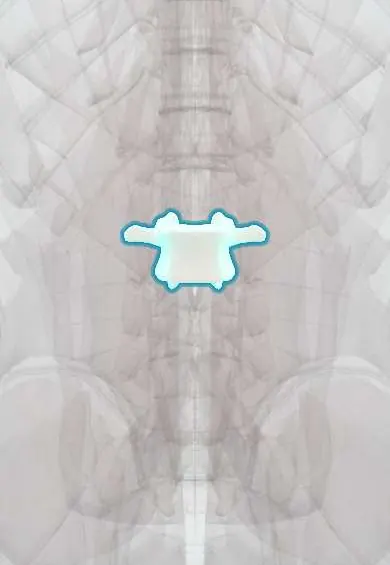
L2 vertebra !
L2 is the second lumbar (lower-back) vertebra; its large body carries the weight of the upper body and lets the lower back bend.
Official name: L2
Opens in the Durgical app, no sign-up needed.
What it is
The L2 lower-back vertebra, with a large body built to carry weight.
What it does
Carries the weight of the upper body and, with the disc and facet joints, lets the lower back bend forward and back.
Key facts
- Joints with
- L1 above and L3 below (intervertebral discs, facet joints)
- Landmarks
- Large kidney-shaped body; no costal facets or transverse foramina; mammillary processes; lower limit of conus medullaris (varies T12–L3); diaphragm crura attach to L1–L3 bodies
Relations
- In front
- Abdominal aorta with the renal arteries leaving at about L1–L2, the left renal vein crossing in front of it, the inferior vena cava to the right and the cisterna chyli beside the right crus
- Lateral
- Psoas major with the lumbar plexus forming inside it; the sympathetic trunk on the front of the bodies along the edge of the psoas
- Behind
- Quadratus lumborum attaches to the tips of the transverse processes; erector spinae and multifidus under the thoracolumbar fascia
- Contents
- Cauda equina: lumbar and sacral roots floating in the dural sac below the conus medullaris
How to find it
The L2 spine is counted up two from L4, found on the line across the tops of the iliac crests; in a thick back it is easier to count up from the lumbosacral junction on a lateral X-ray.
Examination
- Femoral nerve stretch test: lying prone, the knee is bent and the hip extended; front thigh pain suggests irritation of the L2–L4 roots
On imaging
Lateral and AP lumbar radiographs; level counting starts at the sacrum, and a whole-spine sagittal MRI scout helps when there is a transitional vertebra. At the thoracolumbar junction CT shows burst fractures and fragments pushed back into the canal. Sagittal MRI shows the conus tip, often near L1–L2, and any cord or ligament injury.
Common problems
- Disc degeneration and herniation at L4-L5, the most common level for back and leg pain.
- Degenerative spondylolisthesis: L4 slips forward on L5, often with stenosis (more common in women over 50).
- Spinal stenosis: the canal narrows from disc bulging, facet arthritis and a thick ligamentum flavum.
- Compression fractures from osteoporosis or injury.
How it is diagnosed
X-rays (standing, and bending forward and back) show alignment and slipping. MRI shows discs, nerves and stenosis. CT shows bone detail.
Treatment
- Most back pain improves with staying active, physical therapy and pain medicine.
- Stenosis with leg pain when walking that does not improve: decompression (laminectomy).
- Slipping with instability: decompression plus fusion with screws, rods, a cage and bone graft.
Clinical notes
- Lumbar sympathetic ganglia on the front of the L2–L4 bodies are the target of lumbar sympathetic blocks for some leg pain syndromes and poor limb circulation
- Psoas abscess (from spinal tuberculosis, Crohn disease or the kidney) collects beside these bodies; the hip is held flexed and extending it hurts (psoas sign)
- Upper lumbar fractures from high-energy trauma may come with injuries to the kidneys or the renal vessels in front
Anatomical variants
- The adult conus medullaris ends anywhere from about T12 to L3, most often near the L1–L2 disc; this spread is why needles for lumbar puncture go below L2
Related conditions
Test yourself
Think of the answer, then tap the question to see it.
Why are lumbar punctures made below the L2 vertebra?
Because the conus medullaris can end as low as L2–L3 in some adults.
Which nerve roots do upper lumbar disc herniations at L2–L3 and L3–L4 compress, and what are the signs?
The femoral nerve roots: front thigh pain, weak quadriceps and a reduced knee reflex.
How is the femoral nerve stretch test done, and what does a positive test suggest?
Lying prone, the knee is bent and the hip extended; front thigh pain suggests irritation of the L2–L4 roots.
How does a psoas abscess beside the L2 vertebra show on examination?
The hip is held flexed and extending it hurts (psoas sign).
Educational information, not medical advice. For any symptom, see a health professional.
Its names
- Official Latin (TA2)
- vertebra lumbalis II TA2 1068
- Official Spanish (DeCS)
- Vértebra lumbar L2 DeCS D008159
- English (TA2)
- L2
Related
- L1–L2 disc
- L2–L3 disc
- Lumbar arteries
- Anterior longitudinal ligament
- Diaphragm
- Endplates
- Low-back facet joints
- Interspinales
Sources: Terminologia Anatomica (TA2); DeCS; Anatomy, Back, Lumbar Spine.
Spotted a mistake?We are always improving for the community. Tell us what is wrong and our team will review it.Report a mistake
Representative model, for educational purposes only.