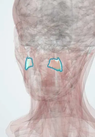
Muscular system · Head and neck
Levator anguli oris !
The levator anguli oris is a small facial muscle running from under the cheek to the corner of the mouth; it lifts the mouth corner.
Opens in the Durgical app, no sign-up needed.
What it is
A small muscle under the cheek to the mouth corner.
What it does
Lifts the corner of the mouth.
Key facts
- Origin
- Canine fossa of the maxilla
- Insertion
- Angle of the mouth
- Action
- Lifts the mouth corner
- Nerve
- Facial nerve (CN VII): zygomatic and buccal branches
- Blood supply
- Facial artery branches
Relations
- Superficial
- Levator labii superioris and zygomaticus minor, with the infraorbital nerve and vessels emerging between them; the facial artery and vein cross it
- Deep
- Canine fossa of the maxilla, below the infraorbital foramen, and the buccinator lower down
- Below
- Modiolus at the angle of the mouth, where it blends with the orbicularis oris and depressor anguli oris
How to find it
Lies deep, under the cheek, between the side of the nose and the mouth corner; it is felt pulling the mouth corner straight up when the person shows the upper canines in a sneer-like smile.
Examination
- Smiling with the upper teeth shown: on a weak side the mouth corner and the nasolabial fold do not rise (zygomatic and buccal branches of the facial nerve)
On imaging
Rarely seen on its own; on ultrasound or MRI it lies deep, arising from the canine fossa just below the infraorbital foramen. Its origin marks the canine space: contrast CT shows an abscess from an upper canine root spreading there, with swelling under the eye and of the nasolabial fold. CT, or cone-beam CT, also shows the tooth and the break in the bone where the infection started.
Clinical notes
- Canine space abscess swells the cheek beside the nose and can close the lower eyelid; the angular and facial veins link this area with the cavernous sinus through the ophthalmic veins
- Infraorbital nerve block, used for upper-lip and cheek procedures, is aimed at the foramen just above its origin
- With the other lip elevators it shapes the nasolabial fold, which flattens in facial palsy
Development
Second pharyngeal arch muscle.
Test yourself
Think of the answer, then tap the question to see it.
Between which muscles does the infraorbital nerve emerge in relation to the levator anguli oris?
Between the levator anguli oris (deep) and the levator labii superioris (superficial).
Where does the levator anguli oris arise, and what is its action?
From the canine fossa of the maxilla; it lifts the mouth corner.
Why is a canine space abscess dangerous?
Pus from an upper canine collects between the levator anguli oris and levator labii superioris; the angular and facial veins link this area with the cavernous sinus.
Educational information, not medical advice. For any symptom, see a health professional.
Its names
- Official Latin (TA2)
- levator anguli oris TA2 2084
- Official Spanish (DeCS)
- Músculo elevador del ángulo de la boca
- English (TA2)
- Levator anguli oris
- Also called
- levator anguli oris muscle · caninus muscle · musculus caninus
Related
Sources: Terminologia Anatomica (TA2); Anatomy, Head and Neck: Levator Anguli Oris Muscle; Anatomy, Head and Neck: Facial Muscles.
Spotted a mistake?We are always improving for the community. Tell us what is wrong and our team will review it.Report a mistake
Representative model, for educational purposes only.