Cardiovascular system · Head and neck
Ophthalmic veins !
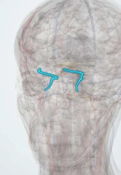
The ophthalmic veins drain the eye socket back into the cavernous sinus; they lack valves, so infection of the face can travel inside the skull.
Official name: Superior ophthalmic vein
Opens in the Durgical app, no sign-up needed.
What it is
Veins of the eye socket draining back into the cavernous sinus.
What it does
Link the face to the cavernous sinus.
Key facts
- Drains
- Orbit and eye; facial vein (angular)
- Into
- Cavernous sinus (inferior also to the pterygoid plexus)
Relations
- Course
- The superior ophthalmic vein starts at the upper inner orbital margin from branches that join the angular vein, runs back under the superior rectus with the ophthalmic artery and leaves through the superior orbital fissure above the common tendinous ring
- Below
- The inferior ophthalmic vein begins as a plexus on the orbital floor, joins the superior vein or the cavernous sinus, and connects through the inferior orbital fissure with the pterygoid plexus
- Behind
- Cavernous sinus, with the internal carotid artery and abducens nerve inside it, and the oculomotor (III), trochlear (IV), ophthalmic (V1) and maxillary (V2) nerves in its lateral wall
How to find it
They cannot be felt. The superior vein begins at the inner upper corner of the orbit from the angular vein and runs back above the optic nerve; for a carotid–cavernous fistula it can be exposed through a small cut in the upper lid crease and entered with a catheter.
Examination
- Listening with the stethoscope bell over the closed eyelid: a whooshing sound in time with the pulse (orbital bruit) supports a carotid–cavernous fistula with flow backward into these veins
On imaging
Contrast CT or MRI: an enlarged superior ophthalmic vein suggests a carotid–cavernous fistula or high pressure in the cavernous sinus; a filling defect in the vein with an enlarged, poorly enhancing cavernous sinus suggests thrombosis. CT also separates preseptal from orbital cellulitis.
Clinical notes
- Preseptal cellulitis stays in front of the orbital septum: red, swollen lids with normal vision and eye movements. Orbital cellulitis lies behind it: pain on eye movement, proptosis, limited movement and sometimes reduced vision
- Orbital cellulitis in children most often spreads from ethmoid sinusitis; through these veins it can reach the cavernous sinus
- The superior ophthalmic vein can serve as an access route for treating a carotid–cavernous fistula from inside the vessel
Related conditions
Test yourself
Think of the answer, then tap the question to see it.
Why can an infection of the face reach the cavernous sinus through the ophthalmic veins?
The ophthalmic veins are valveless, so blood can flow from the facial and angular veins back into the cavernous sinus.
What is the danger triangle of the face and why does it matter?
The area from the corners of the mouth to the bridge of the nose: its veins drain via the angular vein and superior ophthalmic vein to the cavernous sinus.
What does an enlarged superior ophthalmic vein on CT or MRI suggest?
A carotid–cavernous fistula or high pressure in the cavernous sinus.
How is preseptal cellulitis told apart from orbital cellulitis?
Preseptal cellulitis gives swollen lids with normal vision and eye movements; orbital cellulitis adds pain on eye movement, proptosis, limited movement and sometimes reduced vision.
Educational information, not medical advice. For any symptom, see a health professional.
Its names
- Official Latin (TA2)
- vena ophthalmica superior TA2 4887
- Official Spanish (DeCS)
- Venas oftálmicas superior e inferior
- English (TA2)
- Superior ophthalmic vein
Related
Sources: Terminologia Anatomica (TA2); Anatomy, Head and Neck: Eye Ophthalmic Vein.
Spotted a mistake?We are always improving for the community. Tell us what is wrong and our team will review it.Report a mistake
Representative model, for educational purposes only.