Cardiovascular system · Head and neck
Cavernous sinus !
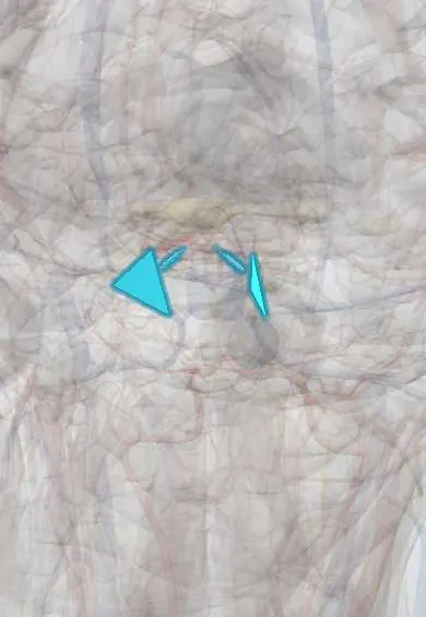
The cavernous sinus is a vein space beside the pituitary through which the internal carotid and eye-moving nerves pass; infection or clots here are dangerous.
Opens in the Durgical app, no sign-up needed.
What it is
A venous space on each side of the sella turcica.
What it does
The internal carotid and nerves III, IV, V1, V2 and VI pass through it; infection or clots here are dangerous.
Key facts
- Drains
- Ophthalmic veins, superficial middle cerebral (sylvian) vein and sphenoparietal sinus, pituitary
- Into
- Superior and inferior petrosal sinuses (contains ICA and CN VI; III, IV, V1, V2 in its wall)
Relations
- Contents
- In the lateral wall, top to bottom: oculomotor nerve, trochlear nerve, ophthalmic nerve (V1), maxillary nerve (V2); inside: the internal carotid artery with its sympathetic plexus, and the abducens nerve lateral to it
- Medial
- Pituitary gland in the sella turcica, and the body of the sphenoid bone with the sphenoid sinus
- Lateral
- Temporal lobe, across the dura
- In front
- Superior orbital fissure, through which it receives the ophthalmic veins
- Behind
- Apex of the petrous temporal bone, where it drains into the petrosal sinuses; the trigeminal ganglion lies just behind and lateral
- Below
- Emissary veins through the skull base link it to the pterygoid plexus
How to find it
It cannot be felt. It lies on each side of the sella turcica and the sphenoid body, from the superior orbital fissure back to the petrous apex; coronal MRI shows it best, and endoscopic surgery reaches its inner wall through the sphenoid sinus.
Examination
- Eye movements, pupils and facial sensation: limited abduction (VI), then ptosis with a dilated pupil (III) and numbness of the forehead and cheek (V1, V2) point to the cavernous sinus; a small pupil (Horner) may appear from the carotid's sympathetic fibres
On imaging
Coronal contrast MRI shows the sinuses enhancing on each side of the pituitary, with the internal carotid as a flow void. Thrombosis gives filling defects, bulging lateral walls and an enlarged superior ophthalmic vein; a fistula gives a dilated superior ophthalmic vein and early filling of the sinus on angiography.
Clinical notes
- Cavernous sinus syndrome: painful eye-movement palsies with numbness of the forehead and cheek; causes include thrombosis, pituitary tumours spreading sideways, meningioma, metastases and granulomatous inflammation (Tolosa–Hunt)
- Cavernous sinus thrombosis: fever, headache, proptosis, chemosis and eye-movement palsies, often becoming bilateral
- An aneurysm of the internal carotid inside the sinus can compress the nerves of the wall without causing subarachnoid haemorrhage
Development
It is one of the few parts of the embryonic primary head vein that survive, the segment beside the trigeminal nerve; the orbital veins drain into it from early on.
Memory aid
«O TOM CAT»: the letters O, T, O, M (oculomotor, trochlear, ophthalmic, maxillary) run down the lateral wall; C and A (carotid, abducens) sit inside.
Related conditions
Test yourself
Think of the answer, then tap the question to see it.
Which nerves run in the lateral wall of the cavernous sinus, from top to bottom?
Oculomotor nerve (III), trochlear nerve (IV), ophthalmic nerve (V1) and maxillary nerve (V2).
Which structures pass through the interior of the cavernous sinus?
The internal carotid artery with its sympathetic plexus, and the abducens nerve (VI) lateral to it.
Which cranial nerve is often affected first in cavernous sinus disease, and what sign results?
The abducens nerve, because it lies free inside the sinus beside the internal carotid: the eye cannot look outward.
By which routes does infection reach the cavernous sinus?
From the facial danger triangle through the valveless facial and ophthalmic veins, from the sphenoid sinus, or from the teeth through the pterygoid plexus.
What is the classic triad of a carotid–cavernous fistula?
Pulsatile exophthalmos, chemosis and an orbital bruit, from arterial blood flooding the cavernous sinus.
Educational information, not medical advice. For any symptom, see a health professional.
Its names
- Official Latin (TA2)
- sinus cavernosus TA2 4860
- Official Spanish (DeCS)
- Seno Cavernoso DeCS D002426
- English (TA2)
- Cavernous sinus
Related
Sources: Terminologia Anatomica (TA2); DeCS; Neuroanatomy, Cavernous Sinus; Anatomy of the Human Body (20th ed.): Development of the Vascular System (Lea & Febiger (Gray's Anatomy, 1918), 1918) · adapted, Public domain.
Spotted a mistake?We are always improving for the community. Tell us what is wrong and our team will review it.Report a mistake
Representative model, for educational purposes only.