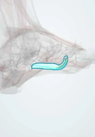
Lumbricals !
The lumbricals of the foot are four small muscles on the toe-flexor tendons in the sole; they help keep the toes straight.
Official name: Lumbrical muscles of foot
Opens in the Durgical app, no sign-up needed.
What it is
Four small, worm-shaped muscles that start on the long toe-flexor tendons.
What it does
Bend the toes at the base while keeping them straight further along, which helps stop them clawing.
Key facts
- Origin
- Four muscles from flexor digitorum longus tendons (first: medial side of first tendon; others: adjacent sides of two tendons)
- Insertion
- Medial side of extensor expansions of toes 2–5
- Action
- Flex MTP and extend interphalangeal joints of toes 2–5
- Nerve
- First: medial plantar nerve; lateral three: lateral plantar nerve, deep branch (S2–S3) (some texts: first two from medial plantar nerve)
- Blood supply
- Lateral plantar artery; plantar metatarsal arteries
Relations
- Course
- Each arises between flexor digitorum longus tendons, runs forward on the medial side of a lesser toe, passes plantar to the deep transverse metatarsal ligament and reaches the extensor expansion
- Superficial
- Flexor digitorum brevis, the plantar fascia and the plantar digital nerves and vessels
- Deep
- Adductor hallucis (third layer) and the interossei (fourth layer)
How to find it
They cannot be felt. In dissection they appear as slender bellies between the flexor digitorum longus tendons once flexor digitorum brevis is reflected.
Examination
- Push-up test: pressing up under the metatarsal heads straightens a flexible claw or hammer toe; if the toe stays bent, the deformity is fixed
On imaging
Too small to assess on radiographs. On ultrasound or MRI they are thin bellies beside the flexor digitorum longus tendons, passing plantar to the deep transverse metatarsal ligament, the level where a Morton neuroma is looked for between the metatarsal heads. In claw toes, radiographs show the joint deformity (extended MTP, flexed interphalangeal joints) rather than the muscles.
Common problems
- Like the other small muscles of the foot, it can weaken with age, nerve problems or tight shoes, which can let the arch drop or the toes change shape.
Clinical notes
- Weakness of the small foot muscles in diabetic neuropathy, hereditary motor and sensory neuropathy or after foot compartment syndrome leads to clawing of the lesser toes
- A common plantar digital nerve runs beside them, plantar to the deep transverse metatarsal ligament; this is where a Morton neuroma forms, most often in the third web space
Memory aid
Hand and foot match: lumbricals arise from the deep long flexor tendons and bend the knuckle while straightening the rest of the finger or toe.
Related conditions
Test yourself
Think of the answer, then tap the question to see it.
What is the nerve supply of the four lumbricals of the foot?
The first: medial plantar nerve; the lateral three: deep branch of the lateral plantar nerve.
What is the action of the lumbricals of the foot?
They flex the MTP joints and extend the interphalangeal joints of toes 2–5, which helps stop clawing.
How do the lumbrical tendons relate to the deep transverse metatarsal ligament, compared with the interossei?
Lumbricals pass plantar to the ligament; the interossei pass dorsal to it.
Educational information, not medical advice. For any symptom, see a health professional.
Its names
- Official Latin (TA2)
- musculi lumbricales pedis TA2 2685
- Official Spanish (DeCS)
- Músculos lumbricales del pie
- English (TA2)
- Lumbrical muscles of foot
- Also called
- lumbricals of foot · lumbrical muscles 1-4 of foot
Related
- Toe flexor tendon
- Medial plantar nerve
- Lateral plantar nerve
- Plantar arteries
- Second metatarsal
- Sesamoids
- Second toe
Sources: Terminologia Anatomica (TA2); Anatomy, Bony Pelvis and Lower Limb, Foot Muscles.
Spotted a mistake?We are always improving for the community. Tell us what is wrong and our team will review it.Report a mistake
Representative model, for educational purposes only.