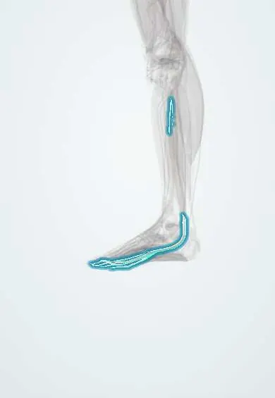
Toe flexor tendon !
The toe flexor tendon runs behind the inner ankle bone, then splits under the foot into tendons that curl the four smaller toes.
Official name: Tendon of flexor digitorum longus
Opens in the Durgical app, no sign-up needed.
What it is
The tendon of the long toe flexor. It runs behind the inner ankle bone and splits under the foot into four tendons to the smaller toes.
What it does
Curls the four smaller toes and helps support the arch. It is often used in tendon transfers for flatfoot.
Key facts
- Attaches
- Behind the medial malleolus, behind the tibialis posterior tendon; crosses superficial to the FHL in the sole (knot of Henry), receives the quadratus plantae, gives origin to the lumbricals and perforates the FDB tendons to the distal phalanges of toes 2–5
- Function
- Flexes toes 2–5 and helps plantarflex and invert; supports the longitudinal arch; a donor tendon in tibialis posterior dysfunction
- Tight in
- Toe extension with ankle dorsiflexion
Relations
- In front
- Tibialis posterior tendon
- Behind
- Posterior tibial artery and veins and the tibial nerve, then the flexor hallucis longus
- Superficial
- Flexor retinaculum; in the sole, the abductor hallucis and the flexor digitorum brevis
- Deep
- Medial side of the sustentaculum tali; in the sole, the quadratus plantae, which inserts on it
How to find it
Behind the medial malleolus, just behind the tibialis posterior; curling the lesser toes against resistance makes it tense, though it is hard to separate from its neighbours by touch.
Examination
- Push-up test: pressing up under the metatarsal heads straightens a flexible claw toe; a toe that stays bent has a fixed deformity
On imaging
Axial MRI and ultrasound show it behind the medial malleolus, second in line: tibialis posterior, flexor digitorum longus, posterior tibial vessels and tibial nerve, then flexor hallucis longus. Its own tears are rare; fluid around it usually means tenosynovitis. Before it is transferred for tibialis posterior dysfunction, MRI or ultrasound confirm that this tendon is healthy.
Clinical notes
- When it is harvested at the knot of Henry, the nearby medial plantar nerve must be protected; the stump is sometimes joined to the flexor hallucis longus to keep some lesser-toe flexion
- The tibial nerve lies right behind it in the tarsal tunnel, where tenosynovitis or a mass can compress the nerve
Anatomical variants
- Tendon slips between the flexor digitorum longus and the flexor hallucis longus in the sole are common, which is why the toes keep some flexion after either is used for a transfer
Memory aid
The 'Dick' in 'Tom, Dick And Very Nervous Harry': second behind the medial malleolus, after the tibialis posterior.
Test yourself
Think of the answer, then tap the question to see it.
Why is the flexor digitorum longus the usual donor tendon in tibialis posterior dysfunction?
It lies beside it, works in phase with it, and its loss costs little because the flexor hallucis longus still flexes the toes.
Where does the flexor digitorum longus cross the flexor hallucis longus?
In the sole, at the knot of Henry, superficial to the flexor hallucis longus.
Which transfer of the flexor digitorum longus corrects flexible claw toes?
The Girdlestone–Taylor transfer, moving it to the top of the proximal phalanx.
Educational information, not medical advice. For any symptom, see a health professional.
Its names
- Official Latin (TA2)
- flexor longus digitorum TA2 2667
- Official Spanish (DeCS)
- Tendón del flexor largo de los dedos DeCS D013710
- English (TA2)
- Tendon of flexor digitorum longus
- Also called
- flexor digitorum longus muscle
Related
- Flexor retinaculum
- Quadratus plantae
- Lumbricals
- Flexor digitorum longus
- Third metatarsal
- Fourth metatarsal
- Fifth metatarsal
- Tibia
Sources: Terminologia Anatomica (TA2); DeCS; Anatomy, Bony Pelvis and Lower Limb: Leg Posterior Compartment.
Spotted a mistake?We are always improving for the community. Tell us what is wrong and our team will review it.Report a mistake
Representative model, for educational purposes only.