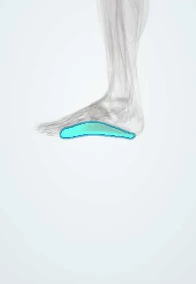
Plantar fascia !
The plantar fascia is a thick band under the sole from the heel to the toes; it supports the arch and can cause heel pain when irritated.
Official name: Plantar aponeurosis
Opens in the Durgical app, no sign-up needed.
What it is
A thick, fibrous band along the sole, from the bottom of the heel bone to the base of the toes.
What it does
Supports the arch like a bowstring. It tightens when the toes bend up, making the foot firm for push-off.
Key facts
- Attaches
- Medial process of calcaneal tuberosity to five slips into plantar plates and fibrous flexor sheaths of toes
- Function
- Supports longitudinal arch; windlass mechanism raises arch on toe dorsiflexion; plantar fasciitis at heel
- Tight in
- Dorsiflexion of toes (especially hallux) with ankle dorsiflexion
Relations
- Course
- From the calcaneal tuberosity it widens forward; near the metatarsal heads each slip splits around the flexor tendons to reach the plantar plates and deep transverse metatarsal ligaments, with fibres to the skin creases
- Superficial
- Heel fat pad, built of fat in fibrous chambers, and the skin of the sole
- Deep
- First layer of sole muscles: flexor digitorum brevis (which arises from its deep surface), abductor hallucis and abductor digiti minimi
- Medial
- Baxter's nerve, passing between the abductor hallucis and the quadratus plantae close to the fascia's origin
How to find it
With the ankle and toes bent up, especially the big toe, the medial edge of the fascia stands out as a taut cord in the sole. The tender point of plantar fasciitis is at the front of the heel pad, over the medial calcaneal tubercle.
Examination
- Windlass test: passive dorsiflexion of the big toe (standing or sitting) reproduces heel pain; specific for plantar fasciitis, though often negative
- Point tenderness at the medial calcaneal tubercle supports plantar fasciitis; pain when the heel is squeezed from both sides suggests a calcaneal stress fracture instead
- Silfverskiöld test: ankle dorsiflexion improves when the knee is bent if the gastrocnemius is tight, a common association
On imaging
Ultrasound shows a thickened, darker origin (more than about 4 mm is the usual cut-off) compared with the other side; MRI shows thickening, partial tears and calcaneal bone oedema. A plantar heel spur on X-ray is common in people without pain and sits mostly in the flexor digitorum brevis origin.
Common problems
- Plantar fasciitis: heel pain, worst with the first steps in the morning or after rest. Common in runners, people who stand all day and people with extra weight.
- Partial tears, sometimes after a sudden push-off.
- Small lumps in the band (plantar fibromatosis).
How it is diagnosed
Usually the history and a physical exam. Ultrasound or MRI can show a thickened fascia. X-rays may show a heel spur, which is often not the cause of the pain.
Treatment
- Most people get better without surgery: stretching the calf and the fascia, supportive shoes or inserts, night splints and activity changes.
- Ongoing pain: physical therapy, injections or shock-wave therapy.
- Surgery to partly release the fascia, for the few people who do not improve after many months.
Clinical notes
- Rupture can follow a sudden push-off and is linked to previous steroid injections; the arch may flatten afterwards
- Release surgery divides only the medial part, because a complete release can flatten the arch and overload the lateral column
- Plantar fibromatosis (Ledderhose disease) forms nodules in the central band and is linked to Dupuytren's disease of the hand
Memory aid
Windlass: "toes up, arch up".
Related conditions
Test yourself
Think of the answer, then tap the question to see it.
What are the attachments of the plantar fascia?
From the medial process of the calcaneal tuberosity to five slips into the plantar plates and fibrous flexor sheaths of the toes.
What is the windlass mechanism of the plantar fascia?
Bending the toes up winds the fascia around the metatarsal heads, raising the arch so the foot becomes a rigid lever for push-off.
Where is the point tenderness of plantar fasciitis, and what does pain on side-to-side heel squeeze suggest instead?
At the medial calcaneal tubercle; pain on squeezing the heel from both sides suggests a calcaneal stress fracture.
Why does plantar fascia release surgery divide only the medial part?
A complete release can flatten the arch and overload the lateral column.
Which nerve entrapment mimics plantar fasciitis heel pain?
Baxter's nerve, the first branch of the lateral plantar nerve, trapped between the abductor hallucis and the quadratus plantae.
Educational information, not medical advice. For any symptom, see a health professional.
Its names
- Official Latin (TA2)
- aponeurosis plantaris TA2 2718
- Official Spanish (DeCS)
- Aponeurosis plantar
- English (TA2)
- Plantar aponeurosis
Related
Sources: Terminologia Anatomica (TA2); Anatomy, Bony Pelvis and Lower Limb, Foot Fascia.
Spotted a mistake?We are always improving for the community. Tell us what is wrong and our team will review it.Report a mistake
Representative model, for educational purposes only.