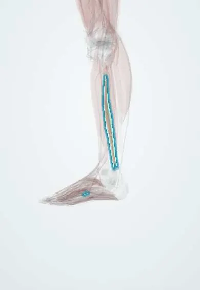
Flexor digitorum longus !
The flexor digitorum longus is a deep calf muscle that curls the four smaller toes; its tendon runs behind the inner ankle.
Opens in the Durgical app, no sign-up needed.
What it is
A deep calf muscle.
What it does
Curls the toes.
Key facts
- Origin
- Medial part of posterior tibia below soleal line
- Insertion
- Bases of distal phalanges of lateral four toes
- Action
- Flexes lateral four toes; plantar flexes ankle; supports longitudinal arch
- Nerve
- Tibial nerve (S2–S3) (some texts: L5–S2)
- Blood supply
- Posterior tibial artery
Relations
- Course
- From the back of the tibia it runs down behind the medial malleolus and under the flexor retinaculum into the sole, where it crosses the flexor hallucis longus tendon and divides into four tendons for toes 2–5
- Deep
- Back of the tibia and tibialis posterior
- Superficial
- Deep transverse fascia and soleus; the posterior tibial vessels and tibial nerve lie between it and flexor hallucis longus
- Lateral
- Tibialis posterior high in the leg, flexor hallucis longus further out
How to find it
Its tendon is felt just behind the tibialis posterior tendon, behind the medial malleolus, when the lesser toes are curled against resistance.
Examination
- Resisted flexion of the tip of a lesser toe while the middle phalanx is held: weakness points to a flexor digitorum longus or tibial nerve lesion (the short flexor acts only up to the middle phalanx)
- Passive extension of the toes is painful in deep posterior compartment syndrome
On imaging
Axial MRI shows it in the deep posterior compartment, on the back of the tibia medial to the tibialis posterior; lower down it crosses superficial to that muscle (crural chiasm). Denervation oedema in it, the tibialis posterior and the flexor hallucis longus points to a tibial nerve lesion. Its tendon is assessed behind the medial malleolus with ultrasound or MRI.
Clinical notes
- Its tendon is the usual replacement for tibialis posterior in reconstruction of flexible adult-acquired flatfoot
- Flexible hammer and claw toes can be treated by moving its tendon slip to the top of the toe (flexor-to-extensor transfer)
- Deep posterior compartment syndrome involves it with tibialis posterior, flexor hallucis longus and the tibial nerve
Anatomical variants
- A tendinous slip linking it with flexor hallucis longus in the sole is common
Development
Its cells gather in the ventral (flexor) muscle mass of the leg bud, which the tibial nerve supplies; the 90° medial turn of the lower limb in week 7 carries this flexor mass round to the back of the leg.
Related conditions
- Plantar Fasciitis
- Tarsal tunnel syndrome
- Medial tibial stress syndrome (shin splints)
- Flatfoot (pes planus)
Test yourself
Think of the answer, then tap the question to see it.
Where does the flexor digitorum longus tendon lie behind the medial malleolus?
Second, between the tibialis posterior tendon in front and the posterior tibial vessels behind.
Which tendon is the usual replacement for the tibialis posterior in flexible adult-acquired flatfoot?
The flexor digitorum longus tendon, transferred to the navicular.
Which sole muscles attach to the flexor digitorum longus tendon?
The quadratus plantae inserts into it and the four lumbricals arise from it, in the second layer of the sole.
Educational information, not medical advice. For any symptom, see a health professional.
Its names
- Official Latin (TA2)
- flexor longus digitorum TA2 2667
- Official Spanish (DeCS)
- Músculo flexor largo de los dedos
- English (TA2)
- Flexor digitorum longus
- Also called
- flexor digitorum longus muscle
Related
Sources: Terminologia Anatomica (TA2); Anatomy, Bony Pelvis and Lower Limb: Leg Posterior Compartment; 8.5 Development of the Appendicular Skeleton (OpenStax (Rice University), 2022) · adapted, CC BY 4.0.
Spotted a mistake?We are always improving for the community. Tell us what is wrong and our team will review it.Report a mistake
Representative model, for educational purposes only.