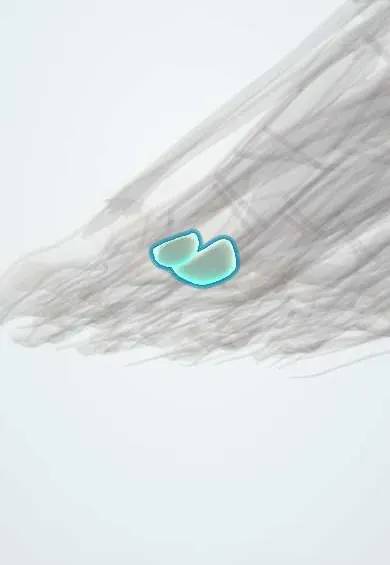
Sesamoids !
The foot sesamoids are two pea-sized bones under the big toe joint, inside a tendon; they work like pulleys and take weight.
Official name: Sesamoid bones of foot
Opens in the Durgical app, no sign-up needed.
What it is
Two small, pea-shaped bones under the head of the first metatarsal, inside the tendons of the flexor hallucis brevis.
What it does
Work like pulleys for the tendons that bend the big toe, and take weight under the ball of the foot.
Key facts
- Joints with
- Plantar surface of first metatarsal head (two grooves)
- Landmarks
- Medial (tibial) and lateral (fibular) sesamoids in flexor hallucis brevis tendons; FHL tendon runs between them; medial larger, may be bipartite
Relations
- Above
- Plantar facets of the first metatarsal head, separated by a central ridge
- Below
- Fat pad and skin of the ball of the foot; the plantar digital nerves of the big toe close to each sesamoid
- Medial
- Abductor hallucis joins the medial head of flexor hallucis brevis at the medial sesamoid
- Lateral
- Adductor hallucis joins the lateral head of flexor hallucis brevis at the lateral sesamoid; deep transverse metatarsal ligament
- In front
- Plantar plate and sesamoid–phalangeal ligaments to the base of the proximal phalanx
How to find it
Felt under the first metatarsal head; dorsiflexing the big toe pulls them forward. Tenderness over one sesamoid that moves with the toe helps tell sesamoid pain from MTP joint pain.
Examination
- Passive dorsiflexion of the big toe while pressing on a sesamoid increases the pain of sesamoiditis or fracture
On imaging
The axial (skyline) sesamoid view shows each sesamoid against the metatarsal head; views of the other foot help tell a bipartite sesamoid from a fracture. MRI shows stress reaction and osteonecrosis; CT or bone scan for hidden fractures.
Common problems
- Sesamoiditis: pain under the big toe joint from overuse, common in dancers and runners.
- Fractures, including stress fractures. Some people are born with a sesamoid in two pieces, which can look like a fracture.
- Pain after turf toe injuries.
How it is diagnosed
A physical exam; X-rays, including special views and a comparison with the other foot; MRI or CT.
Treatment
- Rest, padding to take pressure off, stiff-soled shoes or a boot.
- Physical therapy and a gradual return to activity.
- Surgery to remove or fix a sesamoid that does not heal (uncommon).
Clinical notes
- Removing one sesamoid can unbalance the toe: removing the medial one favours hallux valgus, the lateral one hallux varus
- After turf toe, sesamoids drawn back towards the heel on X-ray suggest a plantar plate rupture
- Osteonecrosis shows as fragmentation and sclerosis of one sesamoid, usually in young women
Anatomical variants
- Sesamoids are also often found at the big toe's interphalangeal joint and under other metatarsal heads, most commonly the fifth
- The os peroneum in the fibularis longus tendon is another foot sesamoid; its frequency varies between populations
Development
Ossify in late childhood, often from more than one centre, which explains bipartite and multipartite sesamoids.
Related conditions
Test yourself
Think of the answer, then tap the question to see it.
In which tendons do the sesamoids of the big toe lie, and which tendon runs between them?
In the tendons of flexor hallucis brevis; the flexor hallucis longus tendon runs between them.
Which sesamoid of the big toe is injured more often?
The medial (tibial) sesamoid: it is larger and takes more load.
How is a bipartite sesamoid told apart from a fracture on X-ray?
Bipartite pieces have smooth rounded edges and together are larger than normal; a fracture has sharp, irregular edges.
Educational information, not medical advice. For any symptom, see a health professional.
Its names
- Official Latin (TA2)
- ossa sesamoidea pedis TA2 1514
- Official Spanish (DeCS)
- Huesos sesamoideos del pie DeCS D012716
- English (TA2)
- Sesamoid bones of foot
Related
- First metatarsal
- MTP ligaments and plantar plate
- Flexor hallucis brevis
- Big toe flexor tendon
- Big toe
- Adductor hallucis
- Lumbricals
Sources: Terminologia Anatomica (TA2); DeCS; Anatomy, Sesamoid Bones.
Spotted a mistake?We are always improving for the community. Tell us what is wrong and our team will review it.Report a mistake
Representative model, for educational purposes only.