Cardiovascular system · Lower limb
Plantar arteries !
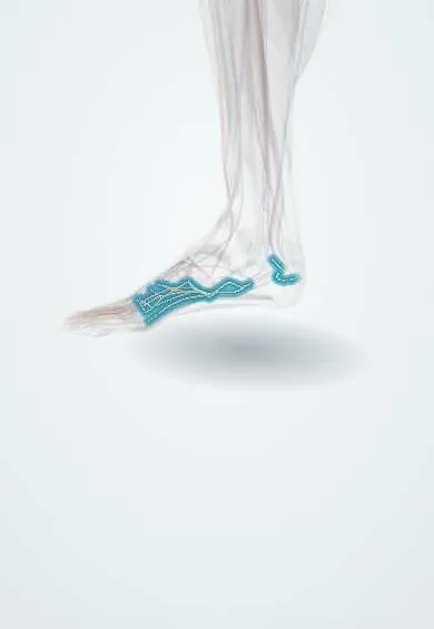
The plantar arteries are branches of the posterior tibial artery that form an arch in the sole and supply the foot.
Official name: Medial plantar artery
Opens in the Durgical app, no sign-up needed.
What it is
The medial and lateral plantar arteries, branches of the posterior tibial artery, and the arch they form in the sole.
What it does
Supply the sole, the small foot muscles and the heel, and connect with the arteries on top of the foot.
Key facts
- Origin
- Posterior tibial artery, dividing under flexor retinaculum and abductor hallucis
- Branches
- Medial plantar (smaller); lateral plantar forms deep plantar arch with plantar metatarsal arteries
- Supplies
- Muscles and skin of sole; toes via plantar digital arteries
Relations
- Course
- Medial: runs forward between the abductor hallucis and flexor digitorum brevis toward the big toe; lateral: crosses the sole obliquely between the flexor digitorum brevis and quadratus plantae to the base of the fifth metatarsal, then turns medially and deep as the plantar arch
- Superficial
- The plantar fascia and the first muscle layer: abductor hallucis, flexor digitorum brevis and abductor digiti minimi
- Deep
- The arch rests on the metatarsal bases and the interossei
How to find it
Not palpable. The bedside check is the posterior tibial pulse just before the division, behind the medial malleolus; a handheld Doppler can follow the lateral plantar artery from there toward the base of the fifth metatarsal.
On imaging
Catheter angiography and CT angiography show the medial and lateral plantar arteries and the deep plantar arch, joined by the dorsalis pedis through the first intermetatarsal space. In diabetic foot disease, angiography shows which arteries still reach the foot and whether the arch is complete before revascularization. Plain radiographs often show calcified artery walls in diabetes.
Clinical notes
- The medial plantar artery flap brings thick, hairless instep skin to rebuild the heel
- Deep wounds and infections of the sole can erode the plantar arch and bleed heavily; it is hard to reach because it lies in the deepest layer
- The sole and heel are posterior tibial territory: heel ulcers heal poorly when the posterior tibial artery and its calcaneal branches are blocked
Anatomical variants
- The relative size of the two contributors to the plantar arch varies: a small lateral plantar artery is offset by a larger deep plantar branch of the dorsalis pedis, and the arch is sometimes incomplete
- A superficial plantar arch, the counterpart of the superficial palmar arch of the hand, is rare (about 5%); the medial plantar artery usually ends in small superficial digital branches instead
Test yourself
Think of the answer, then tap the question to see it.
Where does the posterior tibial artery divide into the plantar arteries?
Under the flexor retinaculum (and abductor hallucis); each branch runs with the plantar nerve of the same name.
How is the deep plantar arch formed?
By the lateral plantar artery, completed medially by the deep plantar branch of the dorsalis pedis artery in the first intermetatarsal space.
Which flap based on a plantar artery is used to rebuild the heel?
The medial plantar artery flap, which brings thick, hairless instep skin.
Educational information, not medical advice. For any symptom, see a health professional.
Its names
- Official Latin (TA2)
- arteria plantaris medialis; arteria plantaris lateralis TA2 4734
- Official Spanish (DeCS)
- Arterias plantares
- English (TA2)
- Medial plantar artery
- Also called
- arteria plantaris tibialis
Related
- Medial plantar nerve
- Lateral plantar nerve
- Toe arteries
- Plantar veins
- Abductor hallucis
- Flexor hallucis brevis
- Adductor hallucis
- Flexor digitorum brevis
Sources: Terminologia Anatomica (TA2); Anatomy, Bony Pelvis and Lower Limb: Foot Arteries; Anatomy of the Human Body (20th ed.): The Arteria Dorsalis Pedis (Lea & Febiger (Gray's Anatomy, 1918), 1918) · adapted, Public domain; Tratado de anatomía humana (9.ª ed.), tomo II, Angiología: aorta descendente, arterias ilíacas y arterias del miembro inferior.
Spotted a mistake?We are always improving for the community. Tell us what is wrong and our team will review it.Report a mistake
Representative model, for educational purposes only.