Joints and ligaments · Upper limb
Lunotriquetral ligament !
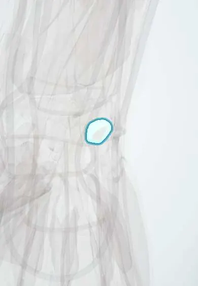
The lunotriquetral ligament joins the lunate and triquetrum in the wrist's first row, so they move as one unit.
Opens in the Durgical app, no sign-up needed.
What it is
A ligament joining the lunate and the triquetrum.
What it does
Keeps the lunate and triquetrum moving as a unit.
Key facts
- Attaches
- Lunate to triquetrum (palmar strongest)
- Function
- Links the lunate and triquetrum
- Tight in
- Radial deviation / pronation loading
Relations
- Above
- TFCC and the ulnocarpal ligaments, with the ulnar head beyond them
- Below
- Hamate and the ulnar side of the capitate, across the midcarpal joint
- In front
- Ulnolunate and ulnotriquetral ligaments and the ulnar arm of the palmar arcuate ligament
- Behind
- Dorsal radiocarpal ligament and dorsal intercarpal ligament, both attached to the triquetrum
How to find it
The LT interval is felt on the back of the wrist just distal to the ulnar head, between the lunate and the triquetrum; it is tender when pressed in LT injuries and is where the ballottement and shear tests are applied.
Examination
- Lunotriquetral ballottement (Reagan): the lunate is fixed between thumb and index of one hand while the triquetrum and pisiform are pushed dorsally and palmarly with the other; pain, crepitus or excess movement suggests an LT tear
- Shear test (Kleinman): the lunate is supported dorsally while the pisiform is pressed dorsally, shearing the joint; pain suggests an LT injury
- LT compression test: pressure on the triquetrum from the ulnar side, just distal to the ulnar styloid, reproduces pain from the LT joint
On imaging
PA view: a step in the first carpal arc (Gilula) between lunate and triquetrum; a visible gap is rare. Lateral view: VISI, with the lunate flexed and a scapholunate angle below about 30°. Wrist arthrography shows contrast passing between the two bones; MR arthrography or arthroscopy confirms the tear.
Clinical notes
- Ulnar impaction can wear through the TFCC disc and then this ligament (Palmer degenerative stages 2D–2E)
- Typical mechanism: a fall on the outstretched hand with the wrist extended and radially deviated, landing on the hypothenar side
- Part of the differential of ulnar-sided wrist pain, with TFCC tears and ECU tendon problems
Anatomical variants
- Lunotriquetral coalition, the commonest carpal coalition, replaces the joint with bone or fibrous tissue; usually found by chance
Development
Carpal coalitions come from incomplete separation of the cartilage precursors of neighbouring carpal bones in the embryo.
Test yourself
Think of the answer, then tap the question to see it.
What happens to the lunate when the lunotriquetral ligament tears?
The lunate follows the scaphoid into flexion, no longer tied to the triquetrum, which tends to extend.
When does a static VISI deformity appear with a lunotriquetral ligament tear?
Only when the dorsal radiocarpal ligament fails as well; isolated tears often leave normal X-rays.
Which ballottement test examines the lunotriquetral ligament?
Reagan's test: the lunate is fixed between thumb and index while the triquetrum and pisiform are pushed dorsally and palmarly; pain or play is positive.
Which stage of a perilunate injury tears the lunotriquetral ligament?
Mayfield stage III, after the scapholunate tear and the capitolunate disruption.
Educational information, not medical advice. For any symptom, see a health professional.
Its names
- Official Latin (TA2)
- ligamentum lunotriquetrum interosseum TA2 1823
- Official Spanish (DeCS)
- Ligamentos semilunopiramidales interóseo y palmar
- English (TA2)
- Lunotriquetral ligament
- Also called
- LT ligament
Related
Sources: Terminologia Anatomica (TA2); Anatomy, Shoulder and Upper Limb, Hand Carpal Bones; Anatomy, Shoulder and Upper Limb, Wrist Joint.
Spotted a mistake?We are always improving for the community. Tell us what is wrong and our team will review it.Report a mistake
Representative model, for educational purposes only.