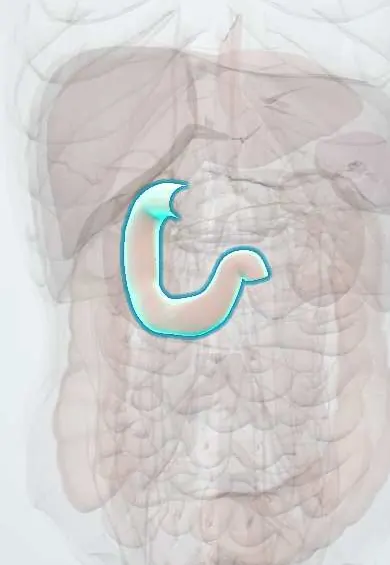
Duodenum !
The duodenum is the C-shaped first part of the small intestine, right after the stomach; bile and pancreatic juice enter it to digest food.
Opens in the Durgical app, no sign-up needed.
What it is
The C-shaped first part of the small intestine, around the head of the pancreas.
What it does
Receives food from the stomach and digestive juices from the liver (bile) and pancreas.
Key facts
- Where
- C-shaped, about 25 cm, around pancreatic head, L1–L3; D1 superior, D2 descending, D3 horizontal, D4 ascending; retroperitoneal except proximal D1
- Blood supply
- Superior pancreaticoduodenal (gastroduodenal, celiac) and inferior pancreaticoduodenal (SMA) arteries; supraduodenal artery for D1
- Nerve
- Vagus and sympathetic fibres via celiac and superior mesenteric plexuses
- Function
- Receives chyme plus bile and pancreatic juice at major papilla (D2); neutralizes acid; secretin and CCK; iron absorption
Relations
- Course
- D1 runs right and back from the pylorus at L1; D2 descends to the right of L1–L3; D3 crosses to the left at L3; D4 rises to the duodenojejunal flexure at L2, held by the suspensory muscle of the duodenum (ligament of Treitz)
- In front
- D1: liver and gallbladder; D2: transverse colon and the root of the transverse mesocolon; D3: superior mesenteric artery and vein and the root of the mesentery
- Behind
- D1: gastroduodenal artery, bile duct and portal vein; D2: right kidney and renal vessels; D3: right ureter, right psoas, inferior vena cava and aorta; D4: left psoas
- Medial
- Head of the pancreas, cradled in its C-shaped curve, with the pancreaticoduodenal arcades between them; the bile duct and main pancreatic duct enter D2 here
- Above
- D1 forms the lower border of the epiploic foramen (of Winslow), with the hepatoduodenal ligament attached to its upper edge
- Lateral
- D2: right colic (hepatic) flexure and the right lobe of the liver
How to find it
Not palpable. At endoscopy the duodenal bulb (cap) lies just past the pylorus; the major papilla is reached on the posteromedial wall of D2 with a side-viewing scope (ERCP). In surgery the Kocher maneuver, incising the peritoneum lateral to D2 and sweeping it medially with the head of the pancreas, exposes its back.
On imaging
A barium meal outlines the triangular duodenal cap; a widened C-loop suggests a mass in the head of the pancreas. CT shows its relation to the superior mesenteric artery and aorta. On a newborn's X-ray, the double-bubble sign (gas in the stomach and proximal duodenum only) points to duodenal atresia or obstruction.
Common problems
- Duodenal ulcer: burning pain, often eased by food; can bleed or perforate.
How it is diagnosed
Examination, X-ray or CT, endoscopy, blood tests.
Treatment
- Depends on the cause: rest and fluids for many obstructions, medicines for Crohn disease, surgery for some.
Clinical notes
- The Kocher maneuver exposes D2, the head of the pancreas and the distal bile duct, and starts a pancreaticoduodenectomy (Whipple procedure)
- Retroperitoneal duodenal injury from handlebar or seat-belt trauma; retroperitoneal air may outline the right kidney on X-ray
- Periampullary tumors (pancreatic head, distal bile duct, ampulla, duodenum) block bile flow early and cause painless jaundice
Anatomical variants
- Pancreas divisum: the dorsal and ventral pancreatic ducts do not join, so most of the gland drains through the minor duodenal papilla, above and in front of the major one; the commonest congenital variant of the pancreas, usually silent
- Annular pancreas: a ring of pancreatic tissue around the second part (D2); uncommon, it can narrow the duodenum and cause vomiting in the newborn or later in life
Development
It comes from the end of the foregut and the start of the midgut, which meet just below the major papilla; that is why both the celiac trunk and the superior mesenteric artery supply it. Failed recanalization causes duodenal atresia, associated with Down syndrome.
Related conditions
- Peptic ulcer disease
- Upper gastrointestinal bleeding
- Coeliac disease
- Infantile hypertrophic pyloric stenosis
- Abetalipoproteinemia (Bassen–Kornzweig syndrome)
- Zinc deficiency (acrodermatitis enteropathica)
- Iron deficiency anaemia
- Iron poisoning
- Zollinger-Ellison syndrome (gastrinoma)
- Functional dyspepsia
- Pancreatic neuroendocrine tumours (VIPoma, glucagonoma, somatostatinoma, non-functioning)
- Giardiasis
- Strongyloidiasis
- Down syndrome (trisomy 21)
- Duodenal and intestinal atresia
- Intestinal malrotation with midgut volvulus
Test yourself
Think of the answer, then tap the question to see it.
Which artery can a posterior ulcer of the first part of the duodenum erode?
The gastroduodenal artery, which runs behind D1, causing heavy bleeding; anterior D1 ulcers tend to perforate instead.
Where does the major duodenal papilla open?
On the posteromedial wall of the second part of the duodenum (D2), where the bile duct and main pancreatic duct open.
Between which vessels does the third part of the duodenum pass?
Between the superior mesenteric artery and vein in front and the aorta and IVC behind; a narrow angle compresses it (SMA syndrome).
What is the Kocher maneuver?
Incising the peritoneum lateral to D2 and sweeping the duodenum and pancreatic head medially, exposing their back and the distal bile duct.
What does the double-bubble sign on a newborn's X-ray suggest?
Duodenal atresia or obstruction: gas only in the stomach and proximal duodenum.
Educational information, not medical advice. For any symptom, see a health professional.
Its names
- Official Latin (TA2)
- duodenum TA2 2944
- Official Spanish (DeCS)
- Duodeno DeCS D004386
- English (TA2)
- Duodenum
- Also called
- intestinum duodenum
Related
Sources: Terminologia Anatomica (TA2); DeCS; Anatomy, Abdomen and Pelvis: Duodenum.
Spotted a mistake?We are always improving for the community. Tell us what is wrong and our team will review it.Report a mistake
Representative model, for educational purposes only.