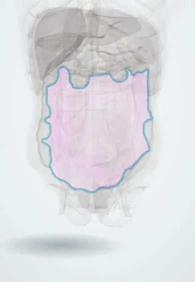
Peritoneum !
The peritoneum is the thin, smooth membrane that lines the inside of the abdomen and covers most of its organs, letting them glide against each other.
Opens in the Durgical app, no sign-up needed.
What it is
The thin, smooth lining of the abdominal cavity, also wrapping many of the organs.
What it does
Lets the organs slide over each other and fights infection. Laparoscopic surgery is done by inflating this space with gas.
Key facts
- Where
- Serous lining of the abdominopelvic cavity: parietal layer on the walls, visceral on organs; posterior parietal layer covers retroperitoneal organs (kidneys, ureters, aorta)
- Function
- Serous fluid lets viscera glide; mesenteries and omenta carry vessels; parietal layer has sharp, well-localized pain, visceral layer poorly localized
Relations
- Superficial
- Extraperitoneal fat and the transversalis fascia separate the parietal peritoneum from the abdominal wall muscles
- Behind
- Retroperitoneum: kidneys, suprarenal glands, ureters, aorta and inferior vena cava, most of the duodenum, the pancreas except its tail, the ascending and descending colon, part of the rectum
- Below
- It drapes over the bladder, uterus and upper rectum, forming the vesicouterine and rectouterine (Douglas) pouches in women and the rectovesical pouch in men
- Contents
- A film of serous fluid; fluid collects in the hepatorenal pouch (Morison pouch, lowest point of the upper abdomen when supine), the subphrenic spaces, the paracolic gutters and the pelvic pouches
How to find it
Paracentesis is usually done in the left lower quadrant, lateral to the rectus sheath and above and medial to the anterior superior iliac spine, to avoid the inferior epigastric artery; or in the midline below the umbilicus through the avascular linea alba, with the bladder emptied.
Examination
- Rebound tenderness (Blumberg sign): pain when pressure is released quickly suggests parietal peritoneal irritation
- Involuntary guarding and rigidity: reflex contraction of the abdominal wall over inflamed parietal peritoneum
- Shifting dullness: the dull area moves when the person turns on their side, which suggests free fluid (ascites); a fluid thrill suggests a large volume
- Cough or jolt tenderness: pain when coughing or with a jarring movement suggests peritonitis
On imaging
An erect chest X-ray shows free air under the diaphragm (pneumoperitoneum) after a perforated viscus; on a supine film, both sides of the bowel wall become visible (Rigler sign). Ultrasound (FAST in trauma) looks for free fluid in Morison pouch, around the spleen and in the pelvis; CT shows ascites, abscesses and peritoneal thickening.
Common problems
- Peritonitis: infection or inflammation, usually from a burst organ (appendix, ulcer); severe pain and a rigid abdomen. An emergency.
How it is diagnosed
Examination, blood tests, CT.
Treatment
- Treat the source (often surgery) and antibiotics.
Clinical notes
- Spontaneous bacterial peritonitis complicates cirrhotic ascites; it is diagnosed on fluid obtained by paracentesis
- After peritonitis, pus collects in the subphrenic and hepatorenal spaces, the lowest points when lying down
- Peritoneal dialysis uses the peritoneum as an exchange membrane
- Tuberculous peritonitis, still seen in Colombia, causes ascites and a doughy abdomen
Development
The peritoneal cavity forms from the intraembryonic coelom; rotation of the gut and fusion of mesenteries with the posterior wall make some organs secondarily retroperitoneal.
Memory aid
Retroperitoneal organs, SAD PUCKER (English mnemonic): Suprarenal glands, Aorta and IVC, Duodenum (parts 2–4), Pancreas (not the tail), Ureters, Colon (ascending and descending), Kidneys, Esophagus, Rectum (part).
Related conditions
- Peritonitis
- Sepsis and septic shock
- Dengue
- Ascites and spontaneous bacterial peritonitis
- Familial Mediterranean fever and other hereditary autoinflammatory syndromes
- Major trauma (polytrauma): ATLS primary survey
- Penetrating trauma (stab and gunshot wounds)
- Blunt abdominal trauma
- Abdominal compartment syndrome
- Intra-abdominal abscess
Test yourself
Think of the answer, then tap the question to see it.
Which abdominal organs are secondarily retroperitoneal?
Most of the duodenum, the pancreas (except the tail), and the ascending and descending colon: they lost their peritoneal covering by fusion to the posterior wall.
What is the only connection between the greater sac and the lesser sac?
The epiploic foramen (of Winslow).
Where is visceral pain from the foregut, midgut and hindgut referred?
Foregut to the epigastrium, midgut around the umbilicus, hindgut to the suprapubic region.
Where does free fluid collect first in the upper abdomen in a supine patient?
The hepatorenal pouch (Morison pouch), the lowest point of the upper abdomen when supine; FAST ultrasound checks it in trauma.
Educational information, not medical advice. For any symptom, see a health professional.
Its names
- Official Latin (TA2)
- peritoneum; peritoneum parietale TA2 3729
- Official Spanish (DeCS)
- Peritoneo DeCS D010537
- English (TA2)
- Peritoneum
- Also called
- peritonaeum
Related
Sources: Terminologia Anatomica (TA2); DeCS; Anatomy, Abdomen and Pelvis, Peritoneum.
Spotted a mistake?We are always improving for the community. Tell us what is wrong and our team will review it.Report a mistake
Representative model, for educational purposes only.