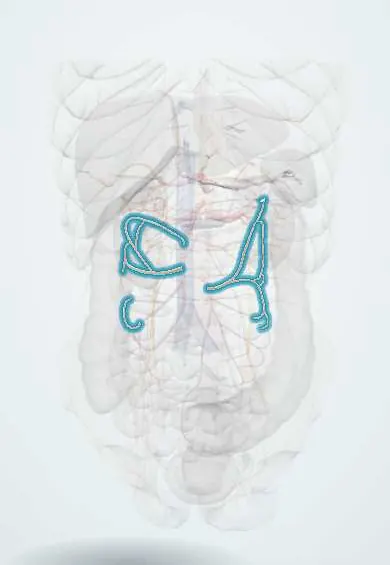
Cardiovascular system · Abdomen
Colic arteries !
The colic arteries feed the colon (large intestine); they link up along its inner edge so the bowel keeps its blood if one branch is lost.
Official name: Right colic artery
Opens in the Durgical app, no sign-up needed.
What it is
The right, middle and left colic arteries, linked along the colon by the marginal artery.
What it does
Supply the colon. A colectomy removes the colon segment fed by the vessel that is tied.
Key facts
- Origin
- Middle and right colic: SMA; left colic: IMA (varies: right colic often from ileocolic or absent)
- Branches
- Ascending and descending branches joining as the marginal artery (of Drummond); inconstant arc of Riolan between middle and left colic
- Supplies
- Right colic: ascending colon; middle colic: transverse colon; left colic: descending colon and splenic flexure
Relations
- Course
- Middle colic: leaves the SMA at the lower border of the pancreas and enters the transverse mesocolon; right colic: runs to the right behind the peritoneum to the ascending colon; left colic: runs left and up from the IMA toward the splenic flexure
- Behind
- Right colic: right gonadal vessels, right ureter and psoas major; left colic: left ureter, left gonadal vessels and psoas major
- Above
- Middle colic: the lower border of the pancreas and the stomach above the transverse mesocolon
How to find it
Found at surgery by lifting the transverse colon and holding its mesocolon up to the light: the middle colic artery shows inside the mesocolon, the ileocolic and right colic arteries run behind the peritoneum toward the right colon, and the left colic artery heads for the descending colon across the front of the left kidney.
On imaging
CT angiography follows each colic artery to its segment before surgery. Mesenteric angiography locates bleeding from diverticula or angiodysplasia and allows embolization.
Common problems
- Mesenteric ischemia: a clot or narrowing starves the gut; severe pain 'out of proportion' to the examination. An emergency.
How it is diagnosed
CT angiography.
Treatment
- Restore flow (catheter or surgery) and remove dead bowel.
Clinical notes
- Colectomies follow these vessels: a right hemicolectomy takes the ileocolic, right colic and right branch of the middle colic arteries; a left hemicolectomy takes the left colic artery or the inferior mesenteric artery
- Ischemic colitis tends to hit the splenic flexure, where the marginal artery between the middle and left colic arteries can be thin or broken (Griffiths point)
- Injury to the middle colic artery during gastric or pancreatic surgery can leave the transverse colon ischemic
Anatomical variants
- The right colic artery is the most variable: it may come from the superior mesenteric artery, the ileocolic artery or the middle colic artery, or be absent
- The middle colic artery may be double, or an accessory middle colic artery may run to the splenic flexure
Development
The ileocolic, right and middle colic arteries belong to the superior mesenteric artery (midgut) and the left colic and sigmoid arteries to the inferior mesenteric artery (hindgut); their meeting point near the splenic flexure marks the old midgut–hindgut border, a watershed zone.
Related conditions
Test yourself
Think of the answer, then tap the question to see it.
Where do the right, middle and left colic arteries arise?
Right and middle colic from the superior mesenteric artery; left colic from the inferior mesenteric artery. The right colic often comes from the ileocolic artery or is absent.
What is the marginal artery (of Drummond)?
A channel joining the colic arteries along the inner border of the colon, so one colic artery can be tied without losing the colon; it can be thin at the splenic flexure.
Why does ischemic colitis tend to hit the splenic flexure?
There the marginal artery between the middle and left colic arteries can be thin or broken (Griffiths point).
Which arteries are taken in a right hemicolectomy?
The ileocolic, right colic and right branch of the middle colic arteries.
What does an enlarged arc of Riolan suggest?
Stenosis of the superior or inferior mesenteric artery; the arc links the middle and left colic arteries.
Educational information, not medical advice. For any symptom, see a health professional.
Its names
- Official Latin (TA2)
- arteria colica dextra; arteria colica media; arteria colica sinistra; arteria marginalis coli TA2 4264
- Official Spanish (DeCS)
- Arterias cólicas
- English (TA2)
- Right colic artery
- Also called
- juxtacolic artery · marginal arcade
Related
Sources: Terminologia Anatomica (TA2); Anatomy, Abdomen and Pelvis: Ileocolic Artery; Anatomy, Abdomen and Pelvis: Superior Mesenteric Artery; Anatomy of the Human Body (20th ed.): The Abdominal Aorta (Lea & Febiger (Gray's Anatomy, 1918), 1918) · adapted, Public domain.
Spotted a mistake?We are always improving for the community. Tell us what is wrong and our team will review it.Report a mistake
Representative model, for educational purposes only.