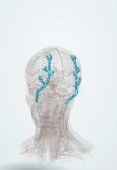
Cardiovascular system · Head and neck
Occipital artery !
The occipital artery is a branch of the external carotid that runs back behind the ear to feed the back of the scalp.
Opens in the Durgical app, no sign-up needed.
What it is
External carotid branches to the back of the scalp and behind the ear.
What it does
Feed the back of the scalp.
Key facts
- Origin
- External carotid
- Branches
- SCM, mastoid, occipital branches (+ posterior auricular artery)
- Supplies
- Back of the scalp, behind the ear
Relations
- Course
- Leaves the back of the external carotid opposite the facial artery, runs back along the lower border of the posterior belly of the digastric, grooves the mastoid part of the temporal bone, crosses the roof of the suboccipital triangle and pierces the fascia at the superior nuchal line to branch over the back of the scalp
- Superficial
- Posterior belly of the digastric, sternocleidomastoid, splenius capitis and longissimus capitis
- Deep
- Near its origin it passes across the internal carotid artery and internal jugular vein, then the vagus and accessory nerves; further back, the superior oblique and semispinalis capitis
- Medial
- On the scalp, the greater occipital nerve
How to find it
The pulse is felt on the superior nuchal line about a third of the way from the external occipital protuberance to the mastoid process; the greater occipital nerve lies just medial to it.
On imaging
Colour Doppler shows it on the back of the scalp beside the greater occipital nerve, a landmark for occipital nerve blocks. CT angiography and catheter angiography show it leaving the back of the external carotid. It often feeds dural arteriovenous fistulas near the transverse and sigmoid sinuses and some posterior fossa tumours, mapped by angiography before embolisation.
Clinical notes
- The greater occipital nerve block uses the pulse of this artery as its landmark, with the injection placed just medial to it
- Scalp wounds bleed heavily: the dense connective tissue of the scalp holds cut vessels open, and the occipital, posterior auricular and superficial temporal arteries anastomose freely
- Neurosurgeons can use it as a donor vessel for bypass to the posterior inferior cerebellar artery
Anatomical variants
- The occipital and posterior auricular arteries may arise from a common trunk
Test yourself
Think of the answer, then tap the question to see it.
Which nerve hooks around the occipital artery just above the carotid bifurcation?
The hypoglossal nerve, which curves forward around the occipital artery or its sternocleidomastoid branch.
How is the occipital artery used as a landmark for a greater occipital nerve block?
Its pulse on the superior nuchal line, a third of the way from the external occipital protuberance to the mastoid; the nerve lies just medial.
Why do scalp wounds over the occipital artery bleed heavily?
Dense scalp tissue holds cut vessels open, and the occipital, posterior auricular and superficial temporal arteries anastomose freely.
Educational information, not medical advice. For any symptom, see a health professional.
Its names
- Official Latin (TA2)
- arteria occipitalis TA2 4398
- Official Spanish (DeCS)
- Arterias occipital y auricular posterior
- English (TA2)
- Occipital artery
Sources: Terminologia Anatomica (TA2); Anatomy, Head and Neck, Occipital Bone, Artery, Vein, and Nerve.
Spotted a mistake?We are always improving for the community. Tell us what is wrong and our team will review it.Report a mistake
Representative model, for educational purposes only.