Skeletal system · Head and neck
Temporal bones !
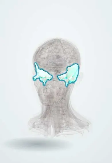
The temporal bones sit at the sides and base of the skull around the ears; they house the hearing and balance organs and form the jaw joint socket.
Official name: Temporal bone
Opens in the Durgical app, no sign-up needed.
What it is
The bones at the sides and base of the skull, around the ears; they contain the middle and inner ear and the mastoid.
What it does
House the hearing and balance organs and form the jaw joint socket.
Key facts
- Joints with
- Parietal, occipital, sphenoid, zygomatic (arch); mandible (TMJ)
- Landmarks
- Squamous, petrous and tympanic parts; mastoid and styloid processes; mandibular fossa; internal and external acoustic meatuses; carotid canal; stylomastoid foramen; articular tubercle; petrotympanic fissure (chorda tympani); arcuate eminence; tegmen tympani; trigeminal impression; jugular fossa
Relations
- Above
- Temporal lobe on the squamous part and the floor of the middle cranial fossa; the trigeminal ganglion in a depression near the petrous apex
- Medial
- Petrous apex next to the cavernous sinus and foramen lacerum; the internal acoustic meatus carries the facial nerve (VII) and the vestibulocochlear nerve (VIII)
- Behind
- Sigmoid sinus in a deep groove on the inner mastoid surface; the cerebellum against the posterior surface of the petrous part
- Below
- Jugular foramen (internal jugular vein, IX, X, XI) and carotid canal under the petrous part; the parotid gland and the facial nerve below the stylomastoid foramen
- Lateral
- Temporalis over the squama; the auricle and ear canal; the mandibular condyle in the mandibular fossa
How to find it
The mastoid process is felt behind the ear lobe and the zygomatic arch in front of the ear. The pterion lies roughly two finger-breadths above the zygomatic arch and a thumb's breadth behind the frontal process of the zygomatic bone.
Examination
- Tuning forks after a head injury: Weber heard louder on the injured side with a negative Rinne there points to conductive loss (blood in the middle ear, broken ossicular chain); Weber going to the other side with a positive Rinne points to sensorineural loss from an inner ear fracture
- Facial movement as soon as possible after the injury (raising the brows, closing the eyes tightly, showing the teeth): weakness of a whole half of the face suggests the facial nerve is hurt in its bony canal, and noting whether it was immediate or delayed guides management
On imaging
High-resolution CT of the temporal bones (thin slices, bone algorithm) shows fractures, the ossicles, the facial canal and the mastoid cells; MRI shows the nerves in the internal acoustic meatus, for example a vestibular schwannoma.
Clinical notes
- An extradural haematoma under the pterion can push the uncus over the tentorium, compressing the oculomotor nerve: a fixed, dilated pupil on the side of the clot
- Acute mastoiditis after otitis media can break through to a postauricular abscess that pushes the ear forward, or spread inward to the sigmoid sinus and meninges
- Gradenigo syndrome: infection of the petrous apex with abducens (VI) palsy, deep facial pain in V1 territory and a discharging ear
- In facial palsy after a temporal bone fracture, immediate onset suggests the nerve is cut; delayed onset suggests swelling, which has a better outlook
Anatomical variants
- Superior semicircular canal dehiscence: the bone over the canal, in the floor of the middle cranial fossa, is very thin or missing; loud sounds or straining can set off vertigo, and some people hear their own pulse or eye movements
- Petrosquamous (Körner) septum: a bony plate that separates superficial from deep mastoid air cells; in mastoid surgery it can be mistaken for the medial limit, leaving deeper diseased cells behind
- Mastoid aeration varies from many large air cells to dense bone with few cells; a poorly aerated mastoid is common in people with long-standing middle ear disease
Development
The mastoid process is barely present at birth and grows over the first years as the sternocleidomastoid pulls on it; the mastoid air cells extend after birth.
Related conditions
Test yourself
Think of the answer, then tap the question to see it.
What are the classic signs of a fracture through the petrous part of the temporal bone?
Bruising over the mastoid (Battle sign), blood behind the eardrum (haemotympanum) and CSF otorrhoea.
Which type of petrous temporal bone fracture more often causes deafness and facial palsy?
The otic capsule violating fracture (older term: transverse), compared with the capsule sparing (longitudinal) type.
Through which foramen of the temporal bone does the facial nerve leave the skull, and why is it vulnerable in infants?
The stylomastoid foramen, between the mastoid and styloid processes; in infants the mastoid is small, so the nerve lies superficial.
What triad defines Gradenigo syndrome of the petrous temporal bone?
Infection of the petrous apex with abducens (VI) palsy, deep facial pain in V1 territory and a discharging ear.
In facial palsy after a temporal bone fracture, what does the timing of onset suggest?
Immediate onset suggests the facial nerve is cut; delayed onset suggests swelling, which has a better outlook.
Educational information, not medical advice. For any symptom, see a health professional.
Its names
- Official Latin (TA2)
- os temporale TA2 641
- Official Spanish (DeCS)
- Hueso Temporal DeCS D013701
- English (TA2)
- Temporal bone
Related
Sources: Terminologia Anatomica (TA2); DeCS; Anatomy, Head and Neck, Skull; Anatomy, Head and Neck, Temporal Region.
Spotted a mistake?We are always improving for the community. Tell us what is wrong and our team will review it.Report a mistake
Representative model, for educational purposes only.