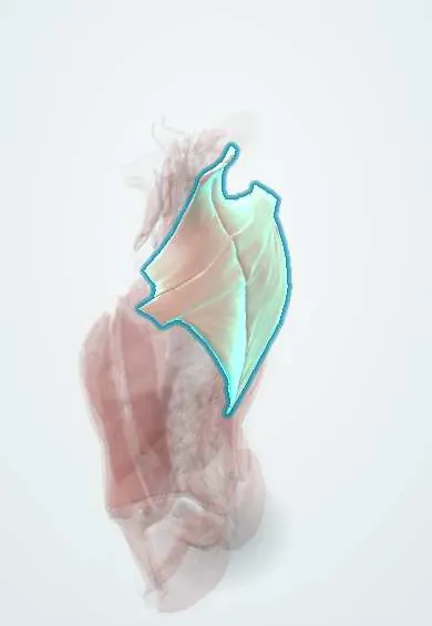
Trapezius !
The trapezius is a large, flat, diamond-shaped muscle covering the back of the neck and upper back; it moves and steadies the shoulder blades.
Official name: Trapezius muscle
Opens in the Durgical app, no sign-up needed.
What it is
A large, flat, diamond-shaped muscle covering the back of the neck and upper back.
What it does
Moves and steadies the shoulder blades; the upper part also extends and tilts the head.
Key facts
- Origin
- Medial superior nuchal line, external occipital protuberance, nuchal ligament, spinous processes of C7–T12
- Insertion
- Lateral third of clavicle, acromion and spine of scapula
- Action
- Upper: elevates scapula, upward rotation; middle: retracts; lower: depresses; upper fibres also extend and tilt the head
- Nerve
- Spinal accessory nerve (CN XI, motor); C3–C4 (proprioception)
- Blood supply
- Transverse cervical artery (superficial branch); occipital, dorsal scapular and posterior intercostal arteries
Relations
- Superficial
- Skin and fat; it is the most superficial muscle of the upper back and neck
- Deep
- Splenius and semispinalis capitis, levator scapulae, rhomboids, supraspinatus and serratus posterior superior; the accessory nerve and superficial branch of the transverse cervical artery on its deep surface
- In front
- Its anterior border bounds the posterior triangle of the neck with sternocleidomastoid and the clavicle
- Below
- Its lower lateral edge, latissimus dorsi and the medial border of the scapula bound the triangle of auscultation
How to find it
The upper fibres stand out when the shoulders are shrugged against resistance; the middle fibres when the shoulder blades are squeezed together; the lower fibres are felt beside the lower thoracic spinous processes when the arm is lifted overhead lying face down.
Examination
- Shrug against resistance tests the accessory nerve: a weak or lower shoulder on one side points to a CN XI lesion, most often in the posterior triangle
- Arm abduction above shoulder height: difficulty with lateral winging of the scapula points to trapezius weakness
On imaging
On axial MRI or CT it is the most superficial muscle of the back of the neck and upper back. After spinal accessory nerve injury, MRI or CT shows trapezius atrophy with fatty replacement, while sternocleidomastoid looks normal if the nerve was cut low in the posterior triangle. Ultrasound shows strains and haematomas and can follow the accessory nerve over levator scapulae.
Clinical notes
- Accessory nerve injury in the posterior triangle (lymph node biopsy, neck dissection) is the commonest cause of trapezius palsy: drooping shoulder, pain and weak overhead reach; sternocleidomastoid is spared because its branch leaves higher up
- Trapezius flaps on the transverse cervical artery are used to cover defects of the back of the neck and the head
- Upper trapezius tension is a frequent finding in tension-type headache and in work-related neck pain
Anatomical variants
- Rarely absent or underdeveloped, in part or completely
Development
Shares an embryonic origin with sternocleidomastoid: one muscle mass in the occipital region, into which the accessory nerve grows before the mass spreads down the back to the shoulder girdle. That shared origin explains the common CN XI supply; the source of the mass (pharyngeal arch or somites) is debated.
Related conditions
Test yourself
Think of the answer, then tap the question to see it.
What nerve supplies the trapezius, and where is it most easily injured?
The spinal accessory nerve (CN XI), injured as it crosses the posterior triangle superficially (lymph node biopsy, neck dissection).
What signs follow paralysis of the trapezius?
A drooping shoulder and lateral scapular winging, most visible on raising the arm sideways, with weak overhead reach.
Which bedside test checks the trapezius and its nerve?
Shrugging against resistance: a weak or lower shoulder on one side points to a CN XI lesion.
Which muscles rotate the scapula upward to lift the arm overhead?
The upper and lower fibres of the trapezius with the serratus anterior.
Why is the sternocleidomastoid spared in accessory nerve injury that paralyses the trapezius?
In posterior triangle injuries, the branch to the sternocleidomastoid has already left higher up.
Educational information, not medical advice. For any symptom, see a health professional.
Its names
- Official Latin (TA2)
- musculus trapezius TA2 2226
- Official Spanish (DeCS)
- Músculo trapecio DeCS D064171
- English (TA2)
- Trapezius muscle
- Also called
- musculus cucullaris
Related
Sources: Terminologia Anatomica (TA2); DeCS; Anatomy, Back, Trapezius; Anatomy of the Human Body (20th ed.): Development of the Muscles (Lea & Febiger (Gray's Anatomy, 1918), 1918) · adapted, Public domain.
Spotted a mistake?We are always improving for the community. Tell us what is wrong and our team will review it.Report a mistake
Representative model, for educational purposes only.