Joints and ligaments · Lower limb
Sole-side midfoot ligaments !
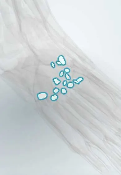
The plantar midfoot ligaments are strong small bands under the midfoot that bind its bones and help hold up the arch.
Official name: Plantar cuneonavicular ligaments
Opens in the Durgical app, no sign-up needed.
What it is
Small ligaments on the sole side of the midfoot, linking the navicular, cuneiforms, cuboid and the bases of the metatarsals.
What it does
Help hold up the arches. They are stronger than the ligaments on top.
Key facts
- Attaches
- Plantar intertarsal, cuneonavicular, cuboideonavicular, intercuneiform and tarsometatarsal ligaments
- Function
- Stronger than dorsal ones; help maintain longitudinal and transverse arches
- Tight in
- Weight-bearing (arch loaded)
Relations
- Superficial
- Fibularis longus tendon crossing obliquely, tibialis posterior tendon slips, then the deep plantar arch with the deep branch of the lateral plantar nerve, the adductor hallucis (oblique head) and the interossei
- Deep
- Plantar surfaces of the navicular, cuneiforms, cuboid and metatarsal bases with their joints
How to find it
Not palpable: they form the deepest layer of the sole, on the bones. In dissection they appear after the four muscle layers are removed, crossed by the fibularis longus tendon and reinforced by slips of the tibialis posterior tendon.
On imaging
Standing lateral X-ray shows plantar gapping or sag at the naviculocuneiform or tarsometatarsal joints when these ligaments fail; MRI shows tears of the plantar Lisfranc ligament.
Clinical notes
- In flatfoot, sag can occur at the midfoot rather than at the talonavicular joint ('midfoot break')
- Charcot neuroarthropathy (most often in diabetes) can collapse the midfoot into a rocker-bottom foot as these ligaments and bones fail
- Bruising in the sole of the midfoot after a twist or crush is a classic sign of a plantar ligament tear in a Lisfranc injury
Test yourself
Think of the answer, then tap the question to see it.
What is the main function of the plantar midfoot ligaments?
Help maintain the longitudinal and transverse arches; they are stronger than the dorsal midfoot ligaments.
Which plantar midfoot ligament marks an unstable Lisfranc injury when torn?
The plantar Lisfranc ligament, from the medial cuneiform to the second and third metatarsal bases.
Which condition, most often in diabetes, collapses the midfoot into a rocker-bottom foot as these ligaments fail?
Charcot neuroarthropathy.
Educational information, not medical advice. For any symptom, see a health professional.
Its names
- Official Latin (TA2)
- ligamenta cuneonavicularia plantaria; ligamenta metatarsea plantaria TA2 1943
- Official Spanish (DeCS)
- Ligamentos plantares del mediopié (cuneonaviculares, intercuneiformes, metatarsianos plantares) DeCS D013640
- English (TA2)
- Plantar cuneonavicular ligaments
- Also called
- ligamenta navicularicuneiformia plantaria
Related
- Lisfranc ligaments
- Top-of-midfoot ligaments
- Cuboid
- Intermediate cuneiform
- Lateral cuneiform
- Medial cuneiform
- First metatarsal
- Second metatarsal
Sources: Terminologia Anatomica (TA2); DeCS; Anatomy, Bony Pelvis and Lower Limb, Foot Joints; Anatomy of the Human Body (20th ed.): Intertarsal Articulations (Lea & Febiger (Gray's Anatomy, 1918), 1918) · adapted, Public domain; Anatomy of the Human Body (20th ed.): Tarsometatarsal Articulations (Lea & Febiger (Gray's Anatomy, 1918), 1918) · adapted, Public domain.
Spotted a mistake?We are always improving for the community. Tell us what is wrong and our team will review it.Report a mistake
Representative model, for educational purposes only.