Joints and ligaments · Lower limb
Top-of-midfoot ligaments !
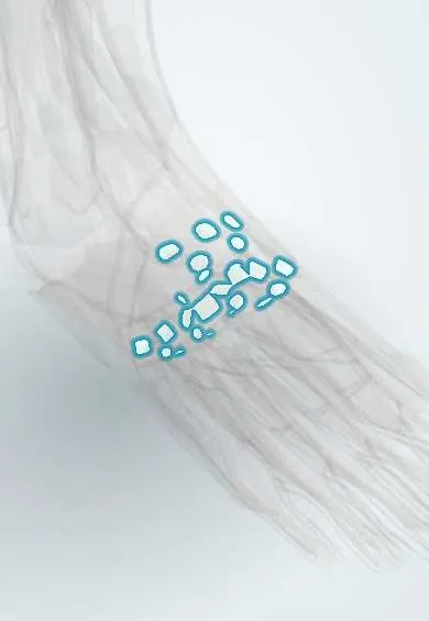
The dorsal midfoot ligaments are many small bands on top of the midfoot linking its bones to each other and to the metatarsals.
Official name: Dorsal cuneonavicular ligaments
Opens in the Durgical app, no sign-up needed.
What it is
Many small ligaments on the top of the midfoot, linking the navicular, cuneiforms, cuboid and the bases of the metatarsals.
What it does
Hold the midfoot bones together as a firm, slightly arched unit.
Key facts
- Attaches
- Dorsal intertarsal, cuneonavicular, cuboideonavicular, intercuneiform and tarsometatarsal ligaments
- Function
- Bind the midfoot bones and metatarsal bases on the dorsal side
- Tight in
- Plantarflexion of midfoot
Relations
- Superficial
- Toe extensor tendons, extensor hallucis longus, the extensor digitorum brevis, the dorsalis pedis artery with the deep fibular nerve, branches of the superficial fibular nerve and the dorsal venous arch
- Deep
- Navicular, cuneiforms, cuboid and metatarsal bases with their intertarsal and tarsometatarsal joints
How to find it
The tuberosity of the fifth metatarsal is easy to feel on the outer border of the foot; from there, the line of tarsometatarsal joints can be followed obliquely across the dorsum. Tenderness along this line after a twist suggests a midfoot sprain.
Examination
- Piano key and pronation–abduction stress tests: pain at a specific joint suggests a dorsal midfoot or Lisfranc sprain
On imaging
Lateral foot X-ray shows dorsal avulsion flakes; standing AP and oblique views check alignment; MRI shows ligament tears and bone bruising.
Common problems
- Sprained in midfoot injuries, including Lisfranc injuries.
Clinical notes
- Dorsal approaches to the midfoot work around the neurovascular bundle that runs between the extensor hallucis longus and the toe extensor tendons
- Dorsal osteophytes of midfoot arthritis ('dorsal bossing') can press on the deep fibular nerve under tight footwear
- In athletes, a midfoot sprain limited to the dorsal ligaments is usually stable; added plantar ligament injury, shown by widening on weight-bearing X-rays, marks an unstable Lisfranc injury
Test yourself
Think of the answer, then tap the question to see it.
How do the dorsal midfoot ligaments compare in strength with the plantar ones, and what follows clinically?
They are thinner and weaker, so midfoot dislocations usually displace toward the top of the foot.
Which bony landmark helps trace the tarsometatarsal joint line across the dorsum?
The tuberosity of the fifth metatarsal: the line runs obliquely from it laterally to the first tarsometatarsal joint medially.
Which nerve can be compressed by «dorsal bossing» of midfoot arthritis under tight footwear?
The deep fibular nerve, pressed by dorsal osteophytes over the midfoot joints.
Educational information, not medical advice. For any symptom, see a health professional.
Its names
- Official Latin (TA2)
- ligamenta cuneonavicularia dorsalia; ligamenta metatarsea dorsalia TA2 1942
- Official Spanish (DeCS)
- Ligamentos dorsales del mediopié (cuneonaviculares, intercuneiformes, metatarsianos dorsales) DeCS D013640
- English (TA2)
- Dorsal cuneonavicular ligaments
- Also called
- ligamenta navicularicuneiformia dorsalia
Related
- Lisfranc ligaments
- Sole-side midfoot ligaments
- Cuboid
- Intermediate cuneiform
- Lateral cuneiform
- Medial cuneiform
- First metatarsal
- Second metatarsal
Sources: Terminologia Anatomica (TA2); DeCS; Anatomy, Bony Pelvis and Lower Limb, Foot Joints.
Spotted a mistake?We are always improving for the community. Tell us what is wrong and our team will review it.Report a mistake
Representative model, for educational purposes only.