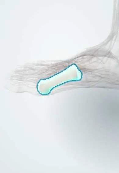
First metatarsal !
The first metatarsal is the short, thick foot bone behind the big toe; it carries the most weight when pushing off.
Official name: First
Opens in the Durgical app, no sign-up needed.
What it is
The short, thick bone behind the big toe. Its head rests on two small sesamoid bones.
What it does
Carries a large share of the body's weight when you stand and push off.
Key facts
- Joints with
- Medial cuneiform; proximal phalanx of big toe; two sesamoids
- Landmarks
- Shortest and thickest metatarsal; tuberosity on base (fibularis longus); head with plantar grooves for sesamoids
Relations
- Behind
- Medial cuneiform at the first tarsometatarsal joint
- In front
- Proximal phalanx of the big toe and the sesamoids
- Below
- Fibularis longus inserting on the plantar tuberosity of the base; the flexor hallucis longus tendon between the sesamoids; flexor hallucis brevis
- Medial
- Part of the tibialis anterior insertion at the base; abductor hallucis; the dorsomedial cutaneous nerve of the big toe runs over the bunion area
- Lateral
- First dorsal interosseous and the first web space; the deep plantar artery passes between the first and second bases
- Above
- Extensor hallucis longus tendon
How to find it
The whole medial border is felt from the base, just in front of the joint with the medial cuneiform, to the head, which forms the medial bulge of the ball of the foot; in hallux valgus the head stands out as the bunion.
On imaging
Standing dorsoplantar and lateral views: hallux valgus and intermetatarsal angles, sesamoid position and joint congruence; in hallux rigidus, a dorsal osteophyte and narrowing. An axial sesamoid view shows the sesamoids under the head.
Common problems
- Bunion (hallux valgus): the bone drifts inward and the big toe leans toward the others, leaving a painful bump.
- Arthritis of the big toe joint (hallux rigidus), with stiffness and a bony bump on top.
- Fractures from a crush or a fall.
How it is diagnosed
A physical exam; X-rays taken standing to measure the angles of the bones.
Treatment
- Wider shoes, padding, toe spacers or inserts to ease pain.
- Bunion surgery: cutting and realigning the bone (osteotomy) or fusing a joint.
- Arthritis: removing bone spurs, fusing the joint or, in some cases, a joint replacement or resurfacing.
Clinical notes
- Bunion surgery chooses between distal (chevron) or proximal osteotomies and first tarsometatarsal fusion (Lapidus) according to the size of the deformity
- A short first metatarsal shifts load to the second metatarsal head (transfer metatarsalgia)
- The dorsomedial cutaneous nerve of the big toe can be injured in bunion incisions, leaving numbness
Anatomical variants
- Os intermetatarseum: an accessory ossicle between the bases of the first and second metatarsals, which can be mistaken for a Lisfranc avulsion fragment
Development
Unlike metatarsals 2–5, its secondary centre is at the base, not the head; a small notch or pseudoepiphysis at the head is normal in children.
Related conditions
Test yourself
Think of the answer, then tap the question to see it.
Where is the secondary ossification centre of the first metatarsal, and how does it differ from the others?
At the base, whereas metatarsals 2–5 have it at the head.
What are the normal upper limits of the hallux valgus angle and the first–second intermetatarsal angle?
Hallux valgus angle under about 15°; intermetatarsal angle under about 9° (some texts: 10°), on a standing AP view.
What is transfer metatarsalgia from a short first metatarsal?
Load shifts to the second metatarsal head, causing pain there.
Which nerve can be injured in bunion incisions, leaving numbness?
The dorsomedial cutaneous nerve of the big toe, running over the bunion area.
Educational information, not medical advice. For any symptom, see a health professional.
Its names
- Official Latin (TA2)
- os metatarsi I TA2 1496
- Official Spanish (DeCS)
- Primer metatarsiano DeCS D008682
- English (TA2)
- First
Related
- Medial cuneiform
- Big toe
- Sesamoids
- Abductor hallucis
- Adductor hallucis
- Crural fascia
- Dorsal interossei
- Extensor hallucis brevis
Sources: Terminologia Anatomica (TA2); DeCS; Anatomy, Bony Pelvis and Lower Limb: Metatarsal Bones.
Spotted a mistake?We are always improving for the community. Tell us what is wrong and our team will review it.Report a mistake
Representative model, for educational purposes only.