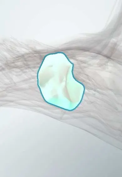
Medial cuneiform !
The medial cuneiform is the largest of three wedge-shaped midfoot bones, behind the big toe's metatarsal.
Official name: Medial cuneiform bone
Opens in the Durgical app, no sign-up needed.
What it is
The largest of three wedge-shaped bones in the midfoot, behind the first metatarsal.
What it does
Their wedge shape helps form the cross arch of the foot. The tibialis anterior tendon attaches here.
Key facts
- Joints with
- Navicular; intermediate cuneiform; metatarsals 1–2
- Landmarks
- Largest cuneiform; tibialis anterior and fibularis longus insert on it; Lisfranc ligament runs from it to base of second metatarsal
Relations
- Behind
- Navicular
- In front
- First metatarsal base at the first tarsometatarsal joint
- Lateral
- Intermediate cuneiform and second metatarsal base, joined by the Lisfranc ligament; the deep plantar artery dives into the sole between the first and second metatarsal bases nearby
- Medial
- Tibialis anterior tendon onto its medial and plantar surface; abductor hallucis below
- Below
- Fibularis longus insertion and a slip of tibialis posterior
How to find it
On the medial border of the foot, between the navicular tuberosity behind and the first metatarsal base in front. With the foot dorsiflexed and inverted, the tibialis anterior tendon is followed down onto it.
Examination
- Piano key test: moving each metatarsal head up and down reproduces midfoot pain in a Lisfranc injury
- Midfoot pain on standing on tiptoe on one leg, or plantar bruising of the midfoot, points to a Lisfranc injury
On imaging
Standing AP view: the medial border of the second metatarsal lines up with the medial border of the intermediate cuneiform, and widening between the first two rays or between the medial and intermediate cuneiforms indicates instability. Comparison standing views, CT or MRI for subtle injuries.
Common problems
- Can be involved in Lisfranc (midfoot) injuries and midfoot arthritis.
Clinical notes
- Lisfranc injuries are often missed at first assessment, leading to collapse of the arch and midfoot arthritis
- Instability of the first tarsometatarsal joint is linked to hallux valgus; fusing it (Lapidus procedure) is one correction
Anatomical variants
- Os intermetatarseum between the first and second metatarsal bases, beside this bone
- Bipartite medial cuneiform, split into dorsal and plantar parts, is rare and can mimic a fracture
Development
Ossifies around age 2, after the lateral cuneiform and before the intermediate one.
Related conditions
Test yourself
Think of the answer, then tap the question to see it.
Which ligament runs from the medial cuneiform to the base of the second metatarsal?
The Lisfranc ligament, the key stabiliser, since no ligament joins the first and second metatarsal bases.
What is the fleck sign on a foot X-ray?
A small avulsed fragment between the first and second metatarsal bases, typical of a Lisfranc injury.
Which two tendons insert on the medial cuneiform from opposite sides?
The tibialis anterior and the fibularis longus, forming a stirrup under the arch.
Educational information, not medical advice. For any symptom, see a health professional.
Its names
- Official Latin (TA2)
- os cuneiforme mediale TA2 1486
- Official Spanish (DeCS)
- Hueso cuneiforme medial DeCS D013639
- English (TA2)
- Medial cuneiform bone
- Also called
- first cuneiform · Cuneiform 1 · os cuneiforme primum
Related
- Navicular
- Intermediate cuneiform
- First metatarsal
- Lisfranc ligaments
- Abductor hallucis
- Extensor hallucis brevis
- Extensor hallucis longus
- Fibular retinacula
Sources: Terminologia Anatomica (TA2); DeCS; Anatomy, Bony Pelvis and Lower Limb: Foot and Ankle.
Spotted a mistake?We are always improving for the community. Tell us what is wrong and our team will review it.Report a mistake
Representative model, for educational purposes only.