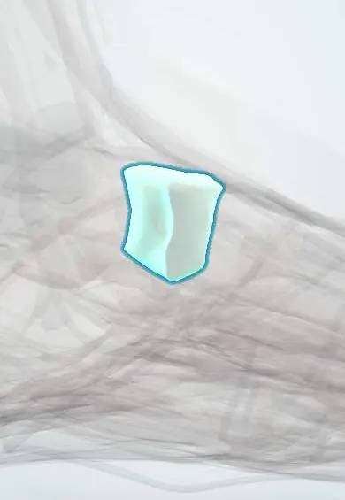
Intermediate cuneiform !
The intermediate cuneiform is the smallest wedge-shaped midfoot bone, behind the second metatarsal.
Official name: Intermediate cuneiform bone
Opens in the Durgical app, no sign-up needed.
What it is
The smallest of three wedge-shaped bones in the midfoot, behind the second metatarsal.
What it does
Their wedge shape helps form the cross arch of the foot. It sits a little back, so the second metatarsal locks in like a keystone.
Key facts
- Joints with
- Navicular; medial and lateral cuneiforms; second metatarsal
- Landmarks
- Smallest cuneiform; set back, forming a recess that locks the base of the second metatarsal; slip of tibialis posterior
Relations
- Behind
- Navicular, middle facet
- In front
- Base of the second metatarsal
- Medial
- Medial cuneiform; the Lisfranc ligament passes deep to the joint between them to the second metatarsal
- Lateral
- Lateral cuneiform
- Above
- The dorsalis pedis artery and deep fibular nerve run over the dorsum near it, lateral to the extensor hallucis longus tendon
- Below
- Plantar ligaments and a slip of tibialis posterior
How to find it
Hard to identify alone; it lies on the dorsum in line with the second metatarsal, roughly where the dorsalis pedis pulse is felt between the extensor hallucis longus and extensor digitorum longus tendons.
Examination
- Midfoot stress (pronation–abduction) test: with the heel held still, twisting the forefoot outward that causes pain at the tarsometatarsal joints suggests a Lisfranc injury
- Piano key test: moving the second metatarsal head up and down reproduces midfoot pain at its base in a Lisfranc injury
- Single-leg heel raise: being unable to rise onto the toes, or pain in the midfoot when trying, suggests an unstable tarsometatarsal complex
On imaging
Standing AP foot view to check its alignment with the second metatarsal base; CT shows the recessed mortise, small avulsions and intercuneiform widening.
Common problems
- Can be involved in Lisfranc (midfoot) injuries and midfoot arthritis.
Clinical notes
- Midfoot arthritis after a Lisfranc injury most often affects the second and third tarsometatarsal joints, the stiff middle column
- Surgical fixation of Lisfranc injuries restores the second metatarsal in its mortise between the cuneiforms
- Bruising on the sole of the midfoot after an injury (plantar ecchymosis sign) points to a Lisfranc injury
Development
The last cuneiform to ossify, around age 3.
Test yourself
Think of the answer, then tap the question to see it.
Why is the second tarsometatarsal joint the least mobile of the foot?
The intermediate cuneiform is set back, so the second metatarsal base sits locked in its recess.
On a standing AP foot view, which alignment is checked at the intermediate cuneiform?
The medial border of the second metatarsal must line up with the medial border of the intermediate cuneiform; any step suggests a Lisfranc injury.
Which is the smallest cuneiform and the last to ossify?
The intermediate cuneiform, ossifying around age 3.
Educational information, not medical advice. For any symptom, see a health professional.
Its names
- Official Latin (TA2)
- os cuneiforme intermedium TA2 1487
- Official Spanish (DeCS)
- Hueso cuneiforme intermedio DeCS D013639
- English (TA2)
- Intermediate cuneiform bone
- Also called
- second cuneiform · Cuneiform 2 · middle cuneiform bone · os cuneiforme secundum
Related
- Navicular
- Medial cuneiform
- Lateral cuneiform
- Second metatarsal
- Extensor hallucis brevis
- Lisfranc ligaments
- Top-of-midfoot ligaments
- Sole-side midfoot ligaments
Sources: Terminologia Anatomica (TA2); DeCS; Anatomy, Bony Pelvis and Lower Limb: Foot and Ankle.
Spotted a mistake?We are always improving for the community. Tell us what is wrong and our team will review it.Report a mistake
Representative model, for educational purposes only.